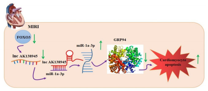
Yanying Wang, Jian Huang, Han Sun, Jie Liu, Yingchun Shao, Manyu Gong, Xuewen Yang, Dongping Liu, Zhuo Wang, Haodong Li, Yanwei Zhang, Xiyang Zhang, Zhiyuan Du, Xiaoping Leng, Lei Jiao, Ying Zhang. Long non-coding RNA-AK138945 regulates myocardial ischemia-reperfusion injury via the miR-1-GRP94 signaling pathway[J]. Frigid Zone Medicine, 2024, 4(1): 31-40. doi: 10.2478/fzm-2024-0004
Citation: Yanying Wang, Jian Huang, Han Sun, Jie Liu, Yingchun Shao, Manyu Gong, Xuewen Yang, Dongping Liu, Zhuo Wang, Haodong Li, Yanwei Zhang, Xiyang Zhang, Zhiyuan Du, Xiaoping Leng, Lei Jiao, Ying Zhang. Long non-coding RNA-AK138945 regulates myocardial ischemia-reperfusion injury via the miR-1-GRP94 signaling pathway[J]. Frigid Zone Medicine, 2024, 4(1): 31-40. doi: 10.2478/fzm-2024-0004

Long non-coding RNA-AK138945 regulates myocardial ischemia-reperfusion injury via the miR-1-GRP94 signaling pathway

doi: 10.2478/fzm-2024-0004
Funds:

the National Natural Science Foundation of China 82370417

the National Natural Science Foundation of China 81970320

the National Natural Science Foundation of China 82270273

the Certificate of China Postdoctoral Science Foundation Grant 2021M693826

the postdoctoral funding from Heilongjiang Province 21042230046

the Hai Yan Youth Fund from Harbin Medical University Cancer Hospital JJQN2021-09

More Information
  •   Objective  Myocardial ischemia-reperfusion injury (MIRI) is one of the leading causes of death from cardiovascular disease in humans, especially in individuals exposed to cold environments. Long non-coding RNAs (lncRNAs) regulate MIRI through multiple mechanisms.This study explored the regulatory effect of lncRNA-AK138945 on myocardial ischemia-reperfusion injury and its mechanism.  Methods  In vivo, 8- to 12-weeks-old C57BL/6 male mice underwent ligation of the left anterior descending coronary artery for 50 minutes followed by reperfusion for 48 hours. In vitro, the primary cultured neonatal mouse ventricular cardiomyocytes (NMVCs) were treated with 100 μmol/L hydrogen peroxide (H2O2). The knockdown of lncRNA-AK138945 was evaluated to detect cardiomyocyte apoptosis, and a glucose-regulated, endoplasmic reticulum stress-related protein 94 (GRP94) inhibitor was used to detect myocardial injury.  Results  We found that the expression level of lncRNA-AK138945 was reduced in MIRI mouse heart tissue and H2O2-treated cardiomyocytes. Moreover, the proportion of apoptosis in cardiomyocytes increased after lncRNA-AK138945 was silenced. The expression level of Bcl2 protein was decreased, and the expression level of Bad, Caspase 9 and Caspase 3 protein was increased. Our further study found that miR-1a-3p is a direct target of lncRNA-AK138945, after lncRNA-AK138945 was silenced in cardiomyocytes, the expression level of miR-1a-3p was increased while the expression level of its downstream protein GRP94 was decreased. Interestingly, treatment with a GRP94 inhibitor (PU-WS13) intensified H2O2-induced cardiomyocyte apoptosis. After overexpression of FOXO3, the expression levels of lncRNA-AK138945 and GRP94 were increased, while the expression levels of miR-1a-3p were decreased.  Conclusion  LncRNA-AK138945 inhibits GRP94 expression by regulating miR-1a-3p, leading to cardiomyocyte apoptosis. The transcription factor Forkhead Box Protein O3 (FOXO3) participates in cardiomyocyte apoptosis induced by endoplasmic reticulum stress through up-regulation of lncRNA-AK138945.


  • Myocardial ischemia-reperfusion injury (MIRI) stands as a significant contributor to the elevated mortality rates of cardiovascular diseases (CVD). Upon its onset, MIRI not only precipitates myocardial arrhythmias but also inflicts irreversible damage upon myocardial cells, ultimately culminating in cardiac dysfunction in afflicted patients[1-3]. Cold weather easily triggers increases in blood pressure and heart rate, potentially leading to myocardial ischemic attacks. Numerous epidemiological studies have highlighted a strong association between low environmental temperature and increased morbidity and mortality associated with CVD[4-7]. Regrettably, effective treatments for MIRI remain absent in clinical practice, and the underlying pathogenesis of MIRI this condition remains elusive. Consequently, the elucidation of MIRI's pathogenesis and the development of novel treatment modalities hold paramount importance.

    Long non-coding RNAs (lncRNAs) exert their regulatory influence on gene expression across various levels of the epigenetic, transcriptional, and post-transcriptional translation processes[8]. They regulate the pathological processes of myocardial fibrosis, myocardial infarction, cardiac hypertrophy, heart failure and other Through interactions with mRNA, DNA, proteins, and microRNAs (miRNAs), they play pivotal roles in modulating the pathological progression of major CVD, such as myocardial fibrosis, myocardial infarction, cardiac hypertrophy, and heart failure[9-11]. Emerging evidence underscores the coordinated involvement of lncRNAs and microRNAs in the regulation of molecules implicated in cardiomyocyte apoptosis. LncRNA-MIRF has been shown tocontribute to cardiomyocyte apoptosis by modulating miR-26a, subsequently promoting the expression of pro-apoptotic protein Bak1[12]. Our previous study has revealed significant dysregulation of LncRNA-AK138945 in a mouse model of cardiac hypertrophy[13], suggesting its potential regulatory significance in CVD. In this study, we explored the effects and underlying mechanisms through which lncRNA-AK138945 modulates MIRI.

    MIRI can precipitate alterations in endoplasmic reticulum (ER) function, leading to the accumulation of unfolded or misfolded proteins and subsequently triggering endoplasmic reticulum stress (ERS). Excessive ERS stands as a pivotal factor in inducing cardiomyocyte apoptosis. Within the process of ERS, GRP94 emerges as a crucial regulator capable of degrading misfolded or unfolded proteins, thereby preserving the stability of the ER environment[14]. Previously, we confirmed that miR-1a-3p can inhibit the expression of GRP94 by targeting its 3'UTR, consequently activating ERS to cause cardiomyocyte apoptosis[15].

    This study elucidated that lncRNA-AK138945 prometed the expression of ERS-related protein GRP94 by targeting miR-1a-3p, thereby playing a role in regulating MIRI-induced cardiomyocyte apoptosis. Therefore, supplementing exogenous or upregulating endogenous lncRNA-AK138945 might emerge as a promising therapeutic strategy for the treatment of MIRI.

    C57BL/6 mice from Liaoning Changsheng Biotechnology Co., Ltd., aged between 8 to 10 weeks and weighing between 22 g and 25 g, were selected for animal experiments. The left anterior descending (LAD) artery coronary artery of the mouse was occluded for 50 minutes using a 7-0 ligature followed by a reperfusion period of 48 hours. (Animal Experimental Ethical Inspection Protocol No. HMUIRB3021619).

    Mice were intraperitoneally injected with 2, 2, 2-tribromoethanol (200 mg/kg; Sigma, St. Louis, Missouri, USA) for anesthesia, and their chests were depilated. The mice were then fixed supine on the operating table, and a proper amount of coupling agent was applied to the chest. The scanning probe was used to record the left ventricular electrocardiogram through the chest wall ultrasound and M-mode echocardiogram. Fractional shortening (FS) and ejection fraction (EF) were determined as the indicators of cardiac function.

    Following the procedure described in our previous study[16], neonatal mouse (C57BL/6) ventricular cells (NMVCs) were treated with hydrogen peroxide (100 μmol/L, Cat#323381, Sigma-Aldrich, USA) for 48 hours to simulate MIRI in vitro. The NMVCs were incubated with GRP94 inhibitor PU-WS13 (5 μmol/L; Cat#58687, MedChemExpress, USA) for 48 hours.

    siRNA targeting lncRNA-AK138945 (5'-GAAGAGGUAUUGAAUG CUA-3') was purchased from Ribobio (Guangzhou, Guangdong Province, China) and transfected into NMVCs following the manufacturer's instructions.

    Cell counting kit-8 (CCK8, CK04-500T, Dojindo, Japan) was used to measure cell viability under different conditions. NMVCs were cultured in 96-well culture clusters (about 1×10⁴ per well), followed by transfection with lncRNA-AK138945 specific siRNA for 48 hours. The CCK8 reagent (20 μL) and DMEM (180 μL) were added to each well and incubated for 1-4 hours. The absorbance was measured at 450 nm using an Infinite m200pro microplate spectrophotometer (Tecan, Salzburg, Austria).

    The cardiomyocytes were seeded on the cover glass of a 24-well culture plate. Forty-eight hours after transfection, live and dead cells were detected using the "Live/Dead Viability/Cytotoxicity Assay Kit" (Invitrogen, Shanghai, China) according to the manufacturer's instructions. A fluorescence microscope (Olympus, Tokyo, Japan) was used to acquire images.

    Terminal deoxynucleotide transferase dUTP nick end labeling (TUNEL) staining was used to evaluate the apoptosis of cultured cardiomyocytes. Briefly, NMVCs cultured on coverslips in 24-well plates were fixed in 4% paraformaldehyde. TUNEL staining was conducted using the in-situ cell death detection kit (Minneapolis, MN, USA) according to the manufacturer's protocol. The numbers of TUNEL-positive cells and the total cells were counted under confocal microscopy.

    Total proteins extracted from NMVCs or mouse left ventricular tissue samples were transferred to a nitrocellulose membrane (Life Science, Überlingen, Germany) using SDS-PAGE (10%-15%). The membrane was sealed with 5% skimmed milk, and incubated overnight at 4℃ with antibodies against GRP94 (Cat#20292, Cell Signaling Technology), Caspase9 (Cat#9504, Cell Signaling Technology), Caspase3 (Cat#9662, Cell Signaling Technology), Bcl2 (Cat#3498, Cell Signaling Technology), Bad (Cat#A19595, ABclonal Technology), or β-actin purchased from ZSGB-BIO (Beijing, China). Then, the membrane was washed with PBST and incubated with a secondary antibody (Alexa Fluor, Molecular Probes, Eugene, OR, USA) in the dark for 50 minutes. Odyssey v1.2 software (LI-COR Biosciences, Lincoln, NB, USA) was used to quantify the immunoblot bands. The results were normalized to the band density of β-actin.

    Total RNA was isolated from myocardial tissue and neonatal cardiomyocytes using TRIzol reagent (Invitrogen, Camarillo, CA, USA). The RNA concentration was measured by a NanoDrop 8000 spectrophotometer (Thermo Scientific, Wilmington, DE, USA). The RNA was reverse transcribed into cDNA using a reverse transcription kit (Dalian, China). Real-time quantitative PCR was performed on the Thermocycler ABI 7500 fast real-time PCR system (Applied Biosystems, Carlsbad, California, USA). The sequences of the primer pairs used in our study are:

    Z FA S 1 - f o r w a r d, 5 ' - A G C G T T T G C T T T G T T C C C - 3 ', r e v e r s e 5 ' - C T C C C T C G AT G C C C T T C T- 3 '; A K 1 3 8 9 4 5 - f o r w a r d, 5 ' - A G C C C A G G A A C A A A T G C A G A A - 3 ', r e v e r s e, 5 ' - T C A A C G T C A C A C T T C AT G AT G G A - 3 '; CDR1as - forward, 5' - TCTGCTCGTCTTCCAACATC - 3', reverse, 5' - AGATCAGCACACTGGAGAC - 3'; β - ACTB - f o r w a r d, 5 ' - A C T G C C G C AT C C T C T T C C T- 3 '; r e v e r s e, 5 ' - T C A A C G T C A C A C T T C AT G AT G G A - 3 '; m i R - 1 a - 3 p - f o r w a r d, 5 ' - G G C G T G G A AT G TA A A G A A - 3 ', r e v e r s e, 5'-CGGCAATTGCACTGGATA-3', miR-1a-3p-RT, 5'-GTCGTATCCAGTGCGTGTCGTGGAGTCGGCAATTGCACTGGATACG ACATACATAC-3'; U6-forward, 5'-GCTTCGGCAGCACATATAC TAAAAT-3', reverse, 5'-CGCTTCACGAATTTGCGTGTCAT-3', U6-RT, 5'-GCTTCGGCAGCACATATACTAAAAT-3'; FOXO3-forward, 5'-CCGTGAGCAAGCCGTGTACTG-3', reverse, 5'-TATCCAGCAGGTCGTCCATGAGG-3'; GRP94-forward, 5' - ATGAAGGCACAAGCATACCAGACG - 3', reverse5' - TCATCTTCCTTAATCCGCCGCAAC-3'.

    HEK293T cells were transfected with miR-1a-3p mimic (20 μmol/L), blank, miR-1a-3p inhibitor, negative control mimic (NC) or negative control inhibitor (NC inhibitor) and plasmid (0.5 μg). Forty-eight hours after transfection, luciferase activity was measured on a fluorometer (Promega, Madison, Wisconsin, USA) using the Dual Luciferase Reporter Assay Kit (Promega, Madison, Wisconsin, USA).

    The experimental data are expressed as mean ± standard error (mean ± SEM) and were statistically analyzed by one-way analysis of variance for multiple group comparisons and t-test for two-group comparisons (GraphPad Prism 8.0 California, United States). P < 0.05 was considered statistically significant.

    To explore the regulatory mechanisms underlying MIRI, we first established a mouse model of MIRI. Comparative analysis revealed a marked reduction in both EF and FS in the MIRI group compared to the Sham group (Fig. 1A). Moreover, the expression levels of pro-apoptotic proteins, including Caspase3, Caspase9, and Bad, were significantly increased in the MIRI group compared to the Sham group (Fig. 1B-D). Subsequently, we assessed the expression levels of circRNA-CDR1as, lncRNA-ZFAS1, and lncRNA-AK138945. We observed a substantial increase in the expression levels of circRNA-CDR1as and lncRNA-ZFAS1 in both the heart tissue of MIRI mice and H2O2-treated cardiomyocytes, whereas the expression level of lncRNA-AK138945 was markedly reduced (Fig. 1E and F). These findings suggest a significant decrease in the expression level of lncRNA-AK138945 during MIRI, indicating its potential regulatory role in his pathological condition.

    Figure  1.  Expression of lncRNA-AK138945 in a mouse model of myocardial ischemia-reperfusion injury (MIRI). (A) Significant decreases in ejection fraction (EF) and fractional shortening (FS) in MIRI mice compared to the control group, indicating the successful establishment of the MIRI model. ** vs. Sham, P < 0.01, Sham, N = 6; MIRI, N = 4. (B-D) Western blot analysis on the expression levels of apoptotic proteins Caspase 9, Caspase 3, and Bad in myocardial tissue. * vs. Sham, P < 0.05; ** vs. Sham, P < 0.01, N = 3-5. (E) qPCR analysis on the expression of lncRNA-AK138945 in neonatal mouse cardiomyocytes after hydrogen peroxide treatment * vs. Control, P < 0.05; ** vs. Control, P < 0.01, N = 3. (F) Changes of lncRNA-AK138945 expression in cardiac tissue of MIRI model. * vs. Sham, P < 0.05; ** vs. Sham, P < 0.01; *** vs. Sham, P < 0.001, N = 3-4.

    To further explore the impact of lncRNA-AK138945 on MIRI, we designed and validated a siRNA targeting lncRNA-AK138945 (siAK138945) and confirmed its efficiency in knockdown (Fig. 2A). Comparative analysis revealed a noticeable reduction in cell viability following the knockdown of lncRNA-AK138945 relative to the control group (Fig. 2B). Moreover, silencing lncRNA-AK138945 increased the ratio of Tunel-positive cells (Fig. 2C), indicating apoptotic cell death. This was further evidenced by the increased cell death rate in siAK138945-treated cardiomyocytes, as revealed by Live/Dead cell staining experiments (Fig. 2D). These findings indicate that knockdown of lncRNA-AK138945 can induce cardiomyocyte apoptosis. To corroborate this inference mechanistically, we examined the effects of lncRNA-AK138945 on apoptosis-related proteins in cardiomyocytes. Noticeably, lncRNA-AK138945 knockdown elevated the levels of pro-apoptotic proteins like Bad, Caspase9, and Caspase3 and reduced the expression of anti-apoptotic protein Bcl-2 (Fig. 3A-D). These data collectively indicate that decreased expression of lncRNA-AK138945 may contribute to MIRI via regulating myocardial cell apoptosis.

    Figure  2.  LncRNA-AK138945 knockdown facilitates cardiomyocyte apoptosis. (A) Verification of lncRNA-AK138945 knockdown efficiency by its siRNA (siAK138945)** vs. Control, P < 0.01; ### vs. +siAK138945, P < 0.001, N = 3. (B) Reduced cardiomyocyte viability by siAK138945, as measured by CCK8 assay. *** vs. Control, P < 0.001; ### vs. +siAK138945, P < 0.001, N = 6. (C) siAK138945 increased cardiomyocyte apoptosis by siAK138945 as unveiled by Tunel staining. ** vs. Control, P < 0.01; ## vs. +siAK138945, P < 0.01, N = 3. (D) siAK138945 increased cardiomyocyte death, as demonstrated by survival/death (Live/Dead) staining. Red fluorescence represents dead cells, and green fluorescence represents live cells. * vs. Control, P < 0.05; # vs. +siAK138945, P < 0.05, N = 3.
    Figure  3.  LncRNA-AK138945 knockdown anomaly alters the expression of apoptosis-related proteins in cardiomyocytes. (A-C) LncRNA-AK138945 knockdown by siAK138945 increases the expression levels of apoptotic proteins Caspase3, Caspase9, and Bad in cardiomyocytes. * vs. Control, P < 0.05; ** vs. Control, P < 0.01; *** vs. Control, P < 0.001; # vs. +siAK138945, P < 0.05; ## vs. +siAK138945, P < 0.01, N = 3-4. (D) siAK138945 significantly reduced the expression of Bcl2. *** vs. Control, P < 0.001; # vs. +siAK138945, P < 0.05, N = 5.

    Subsequently, we endeavored to elucidate why the decreased expression of lncRNA-AK138945 induces myocardial damage. To this end, we conducted an analysis and prediction of microRNAs that can potentially be regulated by lncRNA-AK138945 using a bioinformatics database (http://bibiserv.techfak.uni-bielefeld.de/rnahybrid). Our analysis revealed that lncRNA-AK138945 has the potential to interact with miR-1a-3p, as depicted in Fig. 4A. To confirm the direct interaction between lncRNA-AK138945 and miR-1a-3p, we both cloned the wild-type and mutant miR-1a-3p containing the binding site for lncRNA-AK138945 into a luciferase reporter vector. Then, we co-transfected these constructs with miR-1a-3p, blank, miR-1a-3p inhibitor, NC, and NC inhibitor into HEK293T cells and evaluated the effects of miR-1a-3p on luciferase activity. Comparative analysis revealed a significant reduction in the luciferase activity of lncRNA-AK138945 in the miR-1a-3p group compared to the NC group, and this reduction was restored after mutation of the binding site (Fig. 4B). Moreover, we observed a significant increase in the level of miR-1a-3p following lncRNA-AK138945 knockdown (Fig. 4C). These results collectively indicate that miR-1a-3p is a direct target of lncRNA-AK138945.

    Figure  4.  Experimental verification of miR-1a-3p as a downstream target of lncRNA-AK138945. (A) The predicted binding sites between miR-1a-3p and lncRNA-AK138945. (B) Luciferase reporter gene activity. *** vs. NC, P < 0.001, N = 3. (C) Upregulation of miR-1a-3p expression following lncRNA-AK138945 knockdown. * vs. +siNC, P < 0.05; ** vs. +siNC, P < 0.01, N = 3-6. (D-E) Downregulation of GRP94 expression post-lncRNA-AK138945 knockdown. ** vs. Control, P < 0.01; *** vs. Control, P < 0.001; # vs. +siAK138945, P < 0.05; ## vs. +siAK138945, P < 0.01, N = 5-6.

    We next proceeded to uncover the underlying downstream molecular mechanism of lncRNA-AK138945. Our previous study had already identified miR-1a-3p as a key regulator of MIRI by targeting GRP94[15]. Therefore, we further explored the regulatory relationship between lncRNA-AK138945 and GRP94. As depicted in Fig. 4D and Fig. 4E, lncRNA-AK138945 knockdown markedly reduced the expression of GRP94 at both mRNA and protein levels compared to the siNC group. These findings strongly suggest the existence of the lncRNA-AK138945-miR-1a-3p-GRP94 regulatory pathway during MIRI.

    GRP94 serves as a type of molecular chaperone, actively participating in the ER quality control system and regulating the ER stress signaling pathway. Following myocardial ischemia-reperfusion, there is a notable increase in GRP94 levels[17]. To further investigate the relationship between GRP94 and apoptosis during MIRI, we utilized PU-WS13, a potent GRP94-specific Hsp90 inhibitor with purine scaffolds[18], in our experiments. Upon administration of PU-WS13, there was a significant increase in the expression levels of pro-apoptotic proteins in the H2O2-treated cardiomyocytes, accompanied by a decrease in the expression level of anti-apoptotic proteins (Fig. 5A-D).

    Figure  5.  Inhibition of GRP94 induces cardiomyocyte apoptosis. (A-C) After treatment with a GRP94 inhibitor, the expression of apoptosis-related proteins Caspase3, Caspase9 and Bad was increased. *** vs. Control, P < 0.001; # vs. H2O2, P < 0.05, ## vs. H2O2, P < 0.01, N = 3-6. (D) The expression of anti-apoptotic protein Bcl2 was reduced after treatment with GRP94 inhibitor. ** vs. Control, P < 0.01; # vs. H2O2, P < 0.05, N = 6.

    To further elucidate the upstream mechanism of lncRNA-AK138945 in regulating MIRI, we used the Jaspar database (http://jaspar.genereg.net/) to predict the potential transcription factors of lncRNA-AK138945. As illustrated in Supplementary Material 1, we identified 12 potential FOXO3 recognition/binding sites in the lncRNA-AK138945 sequence, suggesting that FOXO3 may play a role in regulating the lncRNA-AK138945-miR-1-GRP94 signaling pathway. It has been reported that FOXO3 deficiency can lead to increased oxidative damage and decreased myocardial function following acute ischemia-reperfusion injury[19]. Furthermore, our analysis revealed a significant reduction in the expression of FOXO3 during MIRI, further corroborating these findings (Fig. 6A). Subsequently, we constructed a FOXO3 overexpression plasmid and confirmed its efficiency (Fig. 6B). Notably, FOXO3 overexpression significantly increased the expression levels of lncRNA-AK138945 and GRP94, while inhibiting the expression of miR-1a-3p (Fig. 6C-E). This finding suggests that FOXO3 exerts a regulatory effect on the lncRNA-AK138945-miR-1-GRP94 signaling pathway.

    Figure  6.  Effect of transcription factor FOXO3 on the AK138945-miR-1-GRP94 signaling pathway. (A) The expression of FOXO3 in myocardial tissue. *** vs. Sham, P < 0.001, Sham, N = 4; MIRI, N = 4. (B) Verification of FOXO3 overexpression efficiency.*** vs. Control, P < 0.001; # vs. FOXO3, P < 0.05, N = 4. (C-D) The expression levels of lncRNA-AK138945 and GRP94 are up-regulated after FOXO3 overexpression. * vs. Control, P < 0.05; *** vs. Control, P < 0.001; # vs. FOXO3, P < 0.05; ## vs. FOXO3, P < 0.01, N = 6. (E) FOXO3 overexpression downregulates the expression level of miR-1a-3p. ** vs. Control, P < 0.01; # vs. FOXO3, P < 0.05, N = 6.

    Abnormal expression of lncRNA plays a crucial role in CVD, drawing significant attention in recent years due to their regulatory roles in various physiological and pathological processes. Previous studies have documented the involvement of specific lncRNAs in cardiomyocyte apoptosis and MIRI. For instance, lncRNA-6395, recognized as an endogenous proapoptotic factor, regulates cardiomyocyte apoptosis and MIRI by inhibiting the degradation of p53 and promoting its subcellular translocation[20]. Additionally, our group has identified ZFAS1 as elevated during MIRI, attributing to its role in myocardial injury by increasing oxidative stress and cell apoptosis[21]. Furthermore, lncRNA-AK138945 was found to be significantly dysregulated in a mouse model of cardiac hypertrophy in a previous study[11]. In our current work, we took the pioneering step to speculate on its function in MIRI. Surprisingly, findings revealed a marked decrease in the expression of lncRNA-AK138945 in myocardial ischemic cardiac tissue, and lncRNA-AK138945 knockdown resulted in cardiomyocyte injury. However, the involvement of lncRNA participates in other diseases warrants further investigation and discussion.

    Cold-induced vascular dysfunction may further exacerbate the development of CVD[22]. Exposure to cold temperatures can instigate an inflammatory response[23], disrupt the balance between prooxidants and antioxidants[24-25], disturb the Ca2+ homeostasis imbalance, impoact mitochondrial function, induce cell death, and contribute to adverse cardiac hypertrophy, fibrosis, and remodeling[26-30]. Epigenetic changes induced by cold exposure play a crucial role in the pathogenesis of CVD. For instance, cold exposure has been shown to upregulate miR-328, associated with cardiac hypertrophy, while downregulating miR-292-3p expression, linked to aortic endothelial function in mice[31-32]. MiRNAs have emerged as potential targets for the treatment of CVD. For instance, miR-1a-3p inhibits fluoride-induced apoptosis and promotes proliferation of LS8 cells by regulating the MAPK signaling pathway[33]. Moreover, miR-1a-3p has been shown to alleviate symptoms of isoproterenol-induced heart failure in mice by enhancing mitochondrial ND1 and COX1[34]. Our results identified miR-1a-3p as a direct target of lncRNA-AK138945. While regulated by different lncRNAs, miR-1a-3p is likely to be a promising target for the treatment of MIRI. This suggests that the molecular mechanism involving the lncRNA-AK138945-miR-1-GRP94 pathway may be applicable to cold-aggravated MIRI.This study observed detrimental effects on NMVC upon knockdown of LncRNA-AK138945. Further analysis revealed increased expression of miR-1a-3p upon lncRNA-AK138945 knockdown, accompanied by suppressed expression of ERS-related protein GRP94 and cardiomyocyte apoptosis. Consequently, we concluded that lncRNA-AK138945 regulates MIRI through the miR-1-GRP94 signaling pathway. Additionally, FOXO3 was found to exert a regulatory effect on the lncRNA-AK138945-miR-1-GRP94 signaling pathway (Fig. 7).

    Figure  7.  Schematic illustration of the proposed mechanism by which lncRNA-AK138945 regulates myocardial ischemia-reperfusion injury.

    Indeed, despite the significant findings of this study, there are several limitations to that warrant consideration. Firstly, the majority of our experiments were performed at the cellular level, necessitating future investigations to incorporate animal models to better simulate the complexities of in vivo physiological environments. Secondly, further elucidation of the precise mechanism by which AK138945 regulates miR-1a-3p is crucial for a comprehensive understanding of the molecular pathways involved. Finally, while we successfully identified lncRNA-AK138945 as a factor influencing MIRI, continued research efforts are needed to explore its broader implications and potential therapeutic applications. Addressing these limitations will contribute to the advancement of our understanding of MIRI pathogenesis and the development of novel treatment strategies.

  • [1]
    Clark D P, Hanke C W, Swanson N A. Dermal implants: safety of products injected for soft tissue augmentation. J Am Acad Dermatol, 1989(5 Pt 1); 21: 992-998.
    [2]
    Inoue T. Ischemia-reperfusion injury is still a big hurdle to overcome for treatment of acute myocardial infarction. J Cardiol, 2016; 67(4): 305-306. doi: 10.1016/j.jjcc.2015.09.002
    [3]
    Poss J, Desch S, Eitel C, et al. Left ventricular thrombus formation after st-segment-elevation myocardial infarction: insights from a cardiac magnetic resonance multicenter study. Circ Cardiovasc Imaging, 2015; 8(10): e003417. doi: 10.1161/CIRCIMAGING.115.003417
    [4]
    Lv L S, Zhou C L, Jin D H, et al. Impact of ambient temperature on life loss per death from cardiovascular diseases: a multicenter study in central China. Environ Sci Pollut Res Int, 2022; 29(11): 15791-15799. doi: 10.1007/s11356-021-16888-7
    [5]
    Fonseca-Rodriguez O, Sheridan S C, Lundevaller E H, et al. Effect of extreme hot and cold weather on cause-specific hospitalizations in Sweden: A time series analysis. Environ Res, 2021; 193: 110535 doi: 10.1016/j.envres.2020.110535
    [6]
    Hu J, Hou Z, Xu Y, et al. Life loss of cardiovascular diseases per death attributable to ambient temperature: A national time series analysis based on 364 locations in China. Sci Total Environ, 2021; 756: 142614. doi: 10.1016/j.scitotenv.2020.142614
    [7]
    Xu E, Li Y N, Li T T, et al. Association between ambient temperature and ambulance dispatch: a systematic review and meta-analysis. Environ Sci Pollut Res Int, 2022; 29(44): 66335-66347. doi: 10.1007/s11356-022-20508-3
    [8]
    Heo J B, Lee Y S, Sung S. Epigenetic regulation by long noncoding RNAs in plants. Chromosome Res, 2013; 21(6-7): 685-693. doi: 10.1007/s10577-013-9392-6
    [9]
    Zhang Y, Jiao L, Sun L, et al. Lncrna Zfas1 as a Serca2a inhibitor to cause intracellular Ca(2+) overload and contractile dysfunction in a mouse model of myocardial infarction. Circ Res, 2018; 122(10): 1354-1368. doi: 10.1161/CIRCRESAHA.117.312117
    [10]
    Sun F, Zhuang Y, Zhu H, et al. Lncrna Pcfl promotes cardiac fibrosis via mir-378/Grb2 pathway following myocardial infarction. J Mol Cell Cardiol, 2019; 133: 188-198. doi: 10.1016/j.yjmcc.2019.06.011
    [11]
    Cai B, Zhang Y, Zhao Y, et al. Long noncoding RNA-DACH1 (dachshund homolog 1) regulates cardiac function by inhibiting SERCA2a (sarcoplasmic reticulum calciumATPase 2a). Hypertension, 2019; 74(4): 833-842. doi: 10.1161/HYPERTENSIONAHA.119.12998
    [12]
    Su X, Lv L, Li Y, et al. Lncrna Mirf promotes cardiac apoptosis through the miR-26a-Bak1 axis. Mol Ther Nucleic Acids, 2020; 20: 841-850. doi: 10.1016/j.omtn.2020.05.002
    [13]
    Sun L, Zhang Y, Zhuang Y, et al. Expression profile of long non-coding RNAs in a mouse model of cardiac hypertrophy. Int J Cardiol, 2014; 177(1): 73-75. doi: 10.1016/j.ijcard.2014.09.032
    [14]
    Fonseca S G, Urano F, Burcin M, et al. Stress hyperactivation in the beta-cell. Islets, 2010; 2: 1-9. doi: 10.4161/isl.2.1.10456
    [15]
    Zhang Y, Liu X, Zhang L, et al. Metformin protects against H2O2-induced cardiomyocyte injury by inhibiting the miR-1a-3p/GRP94 pathway. Mol Ther Nucleic Acids, 2018; 13: 189-197. doi: 10.1016/j.omtn.2018.09.001
    [16]
    Liu X, Zhang Y, Du W, et al. MiR-223-3p as a novel microrna regulator of expression of voltage-gated K+ channel Kv4.2 in acute myocardial infarction. Cell Physiol Biochem, 2016; 39(1): 102-114. doi: 10.1159/000445609
    [17]
    Martindale J J, Fernandez R, Thuerauf D, et al. Endoplasmic reticulum stress gene induction and protection from ischemia/reperfusion injury in the hearts of transgenic mice with a tamoxifen-regulated form of ATF6. Circ Res, 2006; 98(9): 1186-1193. doi: 10.1161/01.RES.0000220643.65941.8d
    [18]
    Tramentozzi E, Finotti P. Effects of purine-scaffold inhibitors on HUVECs: involvement of the purinergic pathway and interference with ATP. Implications for preventing the adverse effects of extracellular GRP94. Biochem Biophys Rep, 2019; 19: 100661. doi: 10.1016/j.bbrep.2019.100661
    [19]
    Sengupta A, Molkentin J D, Paik J H, et al. Foxo transcription factors promote cardiomyocyte survival upon induction of oxidative stress. J Biol Chem, 2011; 286(9): 7468-7478. doi: 10.1074/jbc.M110.179242
    [20]
    Zhan L F, Zhang Q, Zhao L, et al. LncRNA-6395 promotes myocardial ischemia-reperfusion injury in mice through increasing p53 pathway. Acta Pharmacol Sin, 2022; 43(6): 1383-1394. doi: 10.1038/s41401-021-00767-5
    [21]
    Li M, Jiao L, Shao Y, et al. LncRNA-ZFAS1 promotes myocardial ischemia-reperfusion injury through DNA methylation-mediated Notch1 down-regulation in mice. JACC Basic Transl Sci, 2022; 7(9): 880-895. doi: 10.1016/j.jacbts.2022.06.004
    [22]
    Ebner A, Poitz D M, Alexiou K, et al. Secretion of adiponectin from mouse aorta and its role in cold storage-induced vascular dysfunction. Basic Res Cardiol, 2013; 108(6): 390 doi: 10.1007/s00395-013-0390-9
    [23]
    Xue Y, Yu X, Zhang X, et al. Protective effects of ginsenoside Rc against acute cold exposure-induced myocardial injury in rats. J Food Sci, 2021; 86(7): 3252-3264. doi: 10.1111/1750-3841.15757
    [24]
    Cong P, Liu Y, Liu N, et al. Cold exposure induced oxidative stress and apoptosis in the myocardium by inhibiting the Nrf2-Keap1 signaling pathway. BMC Cardiovasc Disord, 2018; 18(1): 36. doi: 10.1186/s12872-018-0748-x
    [25]
    Park J, Kim S, Kim D H, et al. Whole-body cold tolerance in older Korean female divers "haenyeo" during cold air exposure: effects of repetitive cold exposure and aging. Int J Biometeorol, 2018; 62(4): 543-551. doi: 10.1007/s00484-017-1463-5
    [26]
    Yin Z Q, Ding G B, Chen X, et al. Beclin1 haploinsufficiency rescues low ambient temperature-induced cardiac remodeling and contractile dysfunction through inhibition of ferroptosis and mitochondrial injury. Metabolism, 2020; 113: 154397. doi: 10.1016/j.metabol.2020.154397
    [27]
    Jiang S, Guo R, Zhang Y, et al. Heavy metal scavenger metallothionein mitigates deep hypothermia-induced myocardial contractile anomalies: role of autophagy. Am J Physiol Endocrinol Metab, 2013; 304(1): E74-86. doi: 10.1152/ajpendo.00176.2012
    [28]
    Zhang Y, Li L, Hua Y, et al. Cardiac-specific knockout of ET(A) receptor mitigates low ambient temperature-induced cardiac hypertrophy and contractile dysfunction. J Mol Cell Biol, 2012; 4(2): 97-107. doi: 10.1093/jmcb/mjs002
    [29]
    Zhang Y M, Hu N, Hua Y A, et al. Cardiac over expression of metallothionein rescues cold exposure-induced myocardial contractile dysfunction through attenuation of cardiac fibrosis despite cardiomyocyte mechanical anomalies. Free Radic Biol Med, 2012; 53(2): 194-207. doi: 10.1016/j.freeradbiomed.2012.04.005
    [30]
    Liang J, Yin K, Cao X, et al. Attenuation of low ambient temperature-induced myocardial hypertrophy by atorvastatin via promoting Bcl-2 expression. Cell Physiol Biochem, 2017; 41(1): 286-295. doi: 10.1159/000456111
    [31]
    Yin K, Zhao L, Feng D, et al. Resveratrol attenuated low ambient temperature-induced myocardial hypertrophy via inhibiting cardiomyocyte apoptosis. Cell Physiol Biochem, 2015; 35(6): 2451-2462. doi: 10.1159/000374045
    [32]
    Ebner A, Poitz D M, Alexiou K, et al. Secretion of adiponectin from mouse aorta and its role in cold storage-induced vascular dysfunction. Basic Res Cardiol, 2013; 108(6): 390. doi: 10.1007/s00395-013-0390-9
    [33]
    Chen T, Gu Y, Bai G H, et al. MiR-1a-3p Inhibits apoptosis in fluoride-exposed LS8 cells by targeting Map3k1. Biol Trace Elem Res, 2023.
    [34]
    He R, Ding C, Yin P, et al. MiR-1a-3p mitigates isoproterenol-induced heart failure by enhancing the expression of mitochondrial ND1 and COX1. Exp Cell Res, 2019; 378(1): 87-97. doi: 10.1016/j.yexcr.2019.03.012
  • Relative Articles

    [1]Wen Li, Jia Wang, Yilian Yang, Chunlei Duan, Bing Shao, Mingxiu Zhang, Jiapan Wang, Peifeng Li, Ye Yuan, Yan Zhang, Hongyu Ji, Xingda Li, Zhimin Du. Ethanol extract of cassia seed alleviates metabolic dysfunction-associated steatotic liver disease by acting on multiple lipid metabolism-related pathways[J]. Frigid Zone Medicine, 2024, 4(3): 160-176. doi: 10.1515/fzm-2024-0017
    [2]Cheng Wang, Zican Li, Dongwei Guan, Hongxin Fu, Rennan Feng. Seasonal variation in dietary intake and its association with obesity-related chronic diseases in northeast China[J]. Frigid Zone Medicine, 2024, 4(3): 129-136. doi: 10.1515/fzm-2024-0014
    [3]Jiayue Zhang, Liyao Sun, Xiaohan Yu, Chen Yang, Qi An, Chaoqun Wei, Hongyan Ge. LCN2 aggravates diabetic cataracts by promoting ferroptosis in lens epithelial cells[J]. Frigid Zone Medicine, 2024, 4(3): 177-192. doi: 10.1515/fzm-2024-0018
    [4]Tongzhu Jin, Zhen Ye, Ruonan Fang, Yue Li, Wei Su, Qianqian Wang, Tianyu Li, Hongli Shan, Yanjie Lu, Haihai Liang. Notum protects against myocardial infarction-induced heart dysfunction by alleviating cardiac fibrosis[J]. Frigid Zone Medicine, 2024, 4(1): 41-50. doi: 10.2478/fzm-2024-0005
    [5]Lina Xuan, Huishan Luo, Shu Wang, Guangze Wang, Xingmei Yang, Jun Chen, Jianjun Guo, Xiaomeng Duan, Xiufang Li, Hua Yang, Shengjie Wang, Hailong Zhang, Qingqing Zhang, Shulei Liu, Yongtao She, Kai Kang, Lihua Sun. Circulating CCRR serves as potential novel biomarker for predicting acute myocardial infarction[J]. Frigid Zone Medicine, 2024, 4(3): 137-151. doi: 10.1515/fzm-2024-0015
    [6]Fangfang Bi, Miao Cao, Yuquan Wang, Qingming Pan, Zehong Jing, Danyang Bing, Lifang Lyu, Tong Yu, Tianyu Li, Xuelian Li, Haihai Liang, Hongli Shan, Yuhong Zhou. YBX1 inhibits mitochondrial-mediated apoptosis in ischemic heart through the PI3K/AKT signaling pathway[J]. Frigid Zone Medicine, 2024, 4(1): 51-64. doi: 10.2478/fzm-2024-0006
    [7]Yanan Ni, Dan Liu, Xiaona Zhang, Hong Qiao. Association of point in range with β-cell function and insulin sensitivity of type 2 diabetes mellitus in cold areas[J]. Frigid Zone Medicine, 2023, 3(4): 242-252. doi: 10.2478/fzm-2023-0031
    [8]Manyu Gong, Xuewen Yang, Yaqi Wang, Yanying Wang, Dongping Liu, Haodong Li, Yunmeng Qu, Xiyang Zhang, Yanwei Zhang, Han Sun, Lei Jiao, Ying Zhang. Growth differentiation factor 11 promotes macrophage polarization towards M2 to attenuate myocardial infarction via inhibiting Notch1 signaling pathway[J]. Frigid Zone Medicine, 2023, 3(1): 53-64. doi: 10.2478/fzm-2023-0008
    [9]Juliy M. Perelman, Aleksey B. Pirogov, Anna G. Prikhodko, Victor P. Kolosov. Bronchial inflammatory profile in interferon-gamma-mediated immune response in asthma patients during airway response to cold stimulus[J]. Frigid Zone Medicine, 2022, 2(4): 244-250. doi: 10.2478/fzm-2022-0031
    [10]Yongting Zhao, Xiaofang Zhang, Haihai Liang, Lihong Wang. Antidiabetic agents: Do they hit the right targets?[J]. Frigid Zone Medicine, 2022, 2(4): 225-243. doi: 10.2478/fzm-2022-0030
    [11]Jinxin Liu, Shaohong Fang, Shanjie Wang, Changbin Sun, Rong Sun, Hengxuan Cai, Bo Yu. Effects of intermittent cold-exposure on culprit plaque morphology in ST-segment elevation myocardial infarction patients: a retrospective study based on optical coherence tomography[J]. Frigid Zone Medicine, 2022, 2(2): 82-89. doi: 10.2478/fzm-2022-0011
    [12]Jinglin Wang, Lingfeng Zha. RNA modification by M6A methylation in cardiovascular diseases: Current trends and future directions[J]. Frigid Zone Medicine, 2022, 2(3): 158-177. doi: 10.2478/fzm-2022-0023
    [13]Xiuxiu Wang, Bingjie Hua, Meixi Yu, Shenzhen Liu, Wenya Ma, Fengzhi Ding, Qi Huang, Lai Zhang, Chongwei Bi, Ye Yuan, Mengyu Jin, Tianyi Liu, Ying Yu, Benzhi Cai, Baofeng Yang. Altered expression profile of long non-coding RNAs during heart aging in mice[J]. Frigid Zone Medicine, 2022, 2(2): 109-118. doi: 10.2478/fzm-2022-0015
    [14]Baofeng Yang. Inaugural Editorial[J]. Frigid Zone Medicine, 2021, 1(1): 1-1. doi: 10.2478/fzm-2021-0001
    [15]Yan Feng, Ying Li, Xinling Yang, Limin Han, Luning Wang, Shan Gao, Ruixue Yin, Xue Wang, Jiayang Li, Meiming Liu, Baiyan Li. Direct evidence of VEGF-mediated neuroregulation and afferent explanation of blood pressure dysregulation during angiogenic therapy[J]. Frigid Zone Medicine, 2021, 1(2): 119-126. doi: 10.2478/fzm-2021-0015
    [16]De-an Guo, Wenlong Wei, Changliang Yao, Jianqing Zhang, Qirui Bi. Chinese herbal medicines with beneficial effects[J]. Frigid Zone Medicine, 2021, 1(2): 79-83. doi: 10.2478/fzm-2021-0010
    [17]Ningning Ma, Jing Chen, Jin Ren. Role of microRNAs in the process of metformin treating multiple diseases[J]. Frigid Zone Medicine, 2021, 1(2): 69-78. doi: 10.2478/fzm-2021-0009
    [18]Daba Tolessa Muleta, Xiang Huang, Timur Yagudin, Ying Yang, Jiangang Wang, Xiaoyu Fu, Yue Zhao, Haiyu Gao, Yang Zhou, Zhenwei Pan, Xiangyu Zhong. Ablation of apoptosis-stimulating of p53 protein 1 protects mice from acute hepatic injury and dysfunction via NF-кB pathway in CCl4-induced hepatotoxicity[J]. Frigid Zone Medicine, 2021, 1(1): 53-64. doi: 10.2478/fzm-2021-0007
    [19]Xin Xing, Shiqiang Wang. Mammalian hibernation: a unique model for medical research[J]. Frigid Zone Medicine, 2021, 1(2): 65-68. doi: 10.2478/fzm-2021-0008
    [20]Yanyan Liu, Yahan Yu, Xinyao Wang, Guanqun Liu, Xinda Yin, Yunlong Bai, Zhimin Du. Overexpression of microRNA-135b-5p attenuates acute myocardial infarction injury through its antioxidant and anti-apptotic properties[J]. Frigid Zone Medicine, 2021, 1(2): 85-94. doi: 10.2478/fzm-2021-0011
  • fzm-4-1-31_ESM.pdf
  • 加载中

Catalog

    通讯作者: 陈斌, bchen63@163.com
    • 1. 

      沈阳化工大学材料科学与工程学院 沈阳 110142

    1. 本站搜索
    2. 百度学术搜索
    3. 万方数据库搜索
    4. CNKI搜索

    Figures(7)

    Article Metrics

    Article views (402) PDF downloads(24) Cited by()
    Proportional views
    Related

    /

    DownLoad:  Full-Size Img  PowerPoint
    Return
    Return