Lina Xuan, Huishan Luo, Shu Wang, Guangze Wang, Xingmei Yang, Jun Chen, Jianjun Guo, Xiaomeng Duan, Xiufang Li, Hua Yang, Shengjie Wang, Hailong Zhang, Qingqing Zhang, Shulei Liu, Yongtao She, Kai Kang, Lihua Sun. Circulating CCRR serves as potential novel biomarker for predicting acute myocardial infarction[J]. Frigid Zone Medicine, 2024, 4(3): 137-151. doi: 10.1515/fzm-2024-0015
Citation: Lina Xuan, Huishan Luo, Shu Wang, Guangze Wang, Xingmei Yang, Jun Chen, Jianjun Guo, Xiaomeng Duan, Xiufang Li, Hua Yang, Shengjie Wang, Hailong Zhang, Qingqing Zhang, Shulei Liu, Yongtao She, Kai Kang, Lihua Sun. Circulating CCRR serves as potential novel biomarker for predicting acute myocardial infarction[J]. Frigid Zone Medicine, 2024, 4(3): 137-151. doi: 10.1515/fzm-2024-0015

Circulating CCRR serves as potential novel biomarker for predicting acute myocardial infarction

doi: 10.1515/fzm-2024-0015
Funds:

the Natural Science Foundation of China 81970202

the Natural Science Foundation of China 81903609

Natural Science Foundation of Heilongjiang Province, China LH2022H002

the Outstanding Young Talent Research Fund of College of Pharmacy, Harbin Medical University 2019-JQ-02

2021 (the second batch) Research Funds for affiliated research institutes in Heilongjiang Province CZKYF2021-2-C013

More Information
  • Corresponding author: Kai Kang, E-mail: kangkai1975@sina.com; Lihua Sun, E-mail: sunlihua0219@163.com
  • #These authors contributed equally to this work.

  • Received Date: 2024-04-01
  • Accepted Date: 2024-06-07
  • Available Online: 2024-07-01
  •   Objective   Cold regions exhibit a high prevalence of cardiovascular disease, particularly acute myocardial infarction (AMI), which is one of the leading causes of death associated with cardiovascular conditions. Cardiovascular disease is closely linked to the abnormal expression of long non-coding RNA (lncRNA). This study investigates whether circulating levels of lncRNA cardiac conduction regulatory RNA (CCRR) could serve as a biomarker for AMI.   Materials and methods   We measured circulating CCRR from whole blood samples collected from 68 AMI patients and 69 non-AMI subjects. An AMI model was established using C57BL/6 mice. Quantitative reverse transcription PCR (qRT-PCR) was used to assess CCRR expression. Exosomes were isolated from cardiomyocytes, and their characteristics were evaluated using electron microscope and nanoparticle tracking analysis. The exosome inhibitor GW4869 was employed to examine the effect of exosomal CCRR on cardiac function using echocardiography. Protein expression was detected using Western blot and immunofluorescence staining.   Results   The circulating level of CCRR was significantly higher in AMI patients (1.93 ± 0.13) than in nonAMI subjects (1.00 ± 0.05, P < 0.001). The area under the ROC curve (AUC) of circulating CCRR was 0.821. Similar changes in circulating CCRR levels were consistently observed in an AMI mouse model. Exosomal CCRR derived from hypoxia-induced cardiomyocytes and cardiac tissue after AMI were increased, a change that was reversed by GW4869. Additionally, CCRR-overexpressing exosomes improved cardiac function in AMI.   Conclusion   Circulating lncRNA CCRR is a potential predictor of AMI. Exosomal CCRR plays a role in the communication between the heart and other organs through circulation.


  • The incidence of acute myocardial infarction (AMI) is currently on the rise, contributing significantly to high mortality and morbidity worldwide. In 2019, there were approximately 523 million cases and 18.6 million deaths globally[1]. The Global Burden of Disease Study highlights significant geographical differences in cardiovascular disease mortality rates[2]. An epidemiological study in Asia reveals that mortality from cardiovascular disease is higher in Central and East Asian countries compared to South and Southeast Asian countries[3]. A cohort study on cardiovascular risk factors in China indicates that the prevalence of high cardiovascular disease risk is relatively higher in Northeast China and North China, with the highest rates observed in Northeast China[4]. Northeast China, being a cold region, experiences an increased risk of cardiovascular diseases due to cold-induced changes in the autonomic nervous system, blood pressure, coagulation system, inflammatory response, and oxidative stress[5]. However, previous research on the population risk of cardiovascular disease has limitations, such as limited sample size, narrow distribution of study populations, and a restricted range of risk factors. Early detection is important for the treatment and prognosis of patients with AMI. A reliable noninvasive serum marker is needed for the early identification of AMI patients. Current markers for cardiac muscle necrosis, such as creatine kinase MB (CK-MB), cardiac troponin I (cTnI), Cardiac myosin-binding protein C (cMyBP-C) are useful for detecting AMI[6]. Additionally, biomarkers like soluble suppression of tumorigenicity 2 (sST2), galectin 3 (Gal-3), and bio-ADM correlate with congestion severity and identify risk of death. Growth differentiation factor 15 (GDF-15) might identify a high risk for recurrent cardiovascular events, but these markers have several limitations[7]. Therefore, it is imperative to find more reliable biomarkers especially for the early diagnosis of AMI.

    More recently, long non-coding RNAs (lncRNAs) have emerged as an important component of the gene regulatory network. LncRNAs are a class of non-coding RNAs greater than 200 nucleotides in length, lacking significant open reading frames or protein-coding capacity[8].These transcripts are widely distributed in the cytoplasm and nucleus and are the most abundant class of non-coding RNAs with highly complex functions[9]. LncRNAs participate in numerous physiological and pathological processes, playing regulatory and structural roles in various biological processes[10]. The clinical application value and pathophysiological mechanisms of circulating have been studied lncRNAs as potential biomarkers of cardiovascular diseases[11-13]. Several lncRNAs have been detected in circulation, and we have summarized the potential of these lncRNAs as circulating biomarkers in cardiovascular diseases[14]. Our previous study identified lncRNA-ZAFS1 and lncRNA-CDR1AS as potential diagnostic biomarkers in whole blood of patients with AMI[15]. Early circulating CHAST expression is associated with cardiac contractile function in AMI patients[16]. UCA1 levels in the plasma of AMI patients decrease in the early stage of the disease and increase three days after the onset of myocardial infarction (MI)[17]. Gao et al. showed that HOTAIR is significantly downregulated in the early stage of AMI[18]. MIAT is associated with MI through single nucleotide polymorphism (SNP) association experiment[19]. NEAT1 is part of a molecular circuit involving several chemokines and interleukins that are persistently deregulated post-MI[20]. LIPCAR expression is downregulated after MI and upregulated in late MI[21]. NRF is a potential biomarker of heart failure after AMI[22]. These findings suggest that lncRNAs serve as potential biomarkers for cardiovascular diseases. Our previous study has demonstrated that CCRR is a potential antiarrhythmic target in heart failure, functioning as a signaling mechanism for increased Cx43 protein in the plasma membrane by targeting CIP85[23]. However, the role of CCRR in MI and its potential as a diagnostic marker for MI have not been fully elucidated. Therefore, this study aims to identify the expression changes of CCRR in whole blood after AMI and evaluate its potential as a diagnostic marker for AMI. We collected whole blood samples from AMI patients and non-AMI patients to detect the expression level of CCRR. Additionally, we analyzed the predictive power of lncRNA CCRR for cardiac risk events, correlated its levels with known biomarkers, and assessed the regulatory role of lncRNA CCRR in cardiac function. Ultimately, a comprehensive understanding of how cardiac-derived exosomes promote CCRR trafficking from the myocardium to circulating could provide a basis for novel therapeutic interventions to improve cardiac function following MI.

    This study was conducted in accordance with the Declaration of Helsinki and approved by the Ethnic Committee for the Use of Human Samples at Harbin Medical University. All procedures involving human participants were approved by the Institutional Research Board of Harbin Medical University (No. IRB5003621). Written informed consent was obtained from all study participants. All animal procedures were approved by the Institutional Animal Care and Use Committee at Harbin Medical University.

    The Experimental Animal Center of Harbin Medical University provided male C57BL/6 mice, aged between 8 and 10 weeks and weighing between 20 and 25 grams. All mice were raised in a standard cage with a humidity of 50% ± 20%, a temperature of 23 ± 3℃, and a 12-h light/12-h dark cycle. The mice were kept in the same room with free access to food and water. All experiments complied with the guiding principles for the care and use of laboratory animals at Harbin Medical University and were approved by the Ethics Committee for Animal Experimentation of the School of Pharmacy, Harbin Medical University (No. IRB5003621, No. IRB3001823). Additionally, all animal procedures conformed to the guidelines of Directive 2010/63/EU of the European Parliament on the protection of animals used for scientific purposes.

    Cardiomyocytes were isolated from mice aged 1 to 3 days. In brief, the isolated mouse hearts were washed and chopped in Dulbecco's modified Eagle medium (DMEM) buffer (Biological Industries, Haemek, Israel). The cardiac tissue was digested in trypsin (Beyotime, Shanghai, China) for 12 h and then terminated with DMEM containing 10% Fetal bovine serum (FBS). Subsequently, the cardiac tissue was digested by 0.8 mg/mL type Ⅱ collagenase digestive solution. The resulting cell precipitate was resuspended in DMEM buffer with 10% FBS. For subsequent experiments, cardiomyocytes were collected and inoculated on culture plates for 48 h. To simulate hypoxic conditions, it is essential to maintain the pH balance and metabolic activity of the cardiomyocytes. The cardiomyocytes were cultured under hypoxia conditions at 37℃, with 5% CO2, 1% O2 and 94% N2 for 12 h. The culture medium was collected for exosomes isolation.

    Whole blood sample from 68 AMI patients and 69 non-AMI control subjects were collected from the First Affiliated Hospital of Harbin Medical University (Harbin, China). The samples were obtained via direct venous puncture and collected into the blood collection tubes containing EDTA.

    The mice were anesthetized by intraperitoneal injection of 2, 2, 2-tribromoethanol (Avertin, 0.2 g/kg body weight, Sigma, St. Louis, MO, USA). The thorax was opened at the point of the most pronounced cardiac pulsation to expose the heart. The left anterior descending (LAD) coronary artery was tied with a silk thread, and the chest was subsequently closed. Before surgery, mice in the GW4869 group received an intraperitoneal injection of GW4869 (2.5 mg/kg, MCE). Sham-operated mice served as controls, undergoing the same procedure but without ligation of the LAD coronary artery. For the exosomes administration groups, CCRR-Exos or NC-Exos were injected into the mice via the tail vein. In parallel, sham-operated mice were injected with the same volume of PBS. Model mice with elevated S-T segment in ECGs, confirmed to be suffering from ischemia, were included in the AMI group for follow-up studies.

    Exosomes were isolated by differential centrifugation. The left ventricular tissue of mice, following AMI injury, was removed, washed in PBS, sheared, and digested for 30 min at 37℃ in collagenase type Ⅱ digestion solution (Gibco, USA). Residual tissue and cells were then removed by centrifugation at 1, 000 g for 10 min. The supernatant was collected and centrifuged at 3, 000 g for 10 min, followed by centrifugation at 10, 000 g for 10 min at 4℃. The supernatant was then filtered through a 0.22 μm microporous membrane. The filtered supernatant was centrifuged at 100, 000 g for 2 h at 4 ℃ to pellet all exosomes (Optima L-100XP Ultra centrifuge, Beckman Coulter). After washing with PBS, the exosomes were resuspended in 200 μL PBS for subsequent experiments.

    The freshly isolated cardiomyocyte exosome suspensions were loaded onto 200-mesh carbon-coated Formvar grids for 5 min, and then stained with 2% phosphotungstic acid for 5 min at room temperature. The exosome samples were examined using a transmission electron microscope (TEM; Hitachi, HT7700).

    The particle size distribution of the exosomes was analyzed using nanoparticle tracking analysis (NTA) by the NanoFCM N30E.

    In the exosome uptake experiment, we used the PKH26 Red Fluorescent Cell Linker Mini Kit (Sigma-Aldrich, USA) to label the exosomes. The isolated exosomes were resuspended in PBS, and 100 μL of the exosome suspension was added to 200 μL diluent C. The PKH26 dye was mixed with Diluent C for 5 min at room temperature. The reaction was terminated with 5% BSA in PBS. The labeled exosomes were then isolated using the Ribo™ Exosome Isolation Reagent (Ribobio, China) and resuspended in 100 μL PBS for the uptake experiment. Primary neonatal mouse cardiomyocytes were labeled with α-actinin (A7811, Sigma-Aldrich, USA). The uptake of labeled exosomes by the cardiomyocytes was observed by using laser scanning confocal microscopy.

    The mice were anesthetized by intraperitoneal injection of 2, 2, 2-tribromoethanol (Avertin, 0.2 g/kg body weight, Sigma, St. Louis, MO, USA). The anesthetized mice were then fixed on the workbench and subjected to transthoracic echocardiography using a Vevo 2100 High-Resolution Imaging System (Visual Sonics, Canada). The left ventricular internal diameter diastolic (LVIDd) and the left ventricular internal diameter systolic (LVIDs) were measured in two-dimensional long-axis views. The left ventricular ejection fraction (EF) and fraction shortening (FS) were calculated to assess cardiac function.

    Total RNA was extracted from whole blood using Trizol LS solution (Invitrogen, USA). Exosomes and myocardial tissue from mice in each group were used for RNA extraction with Trizol Regent (Invitrogen, USA). Complementary DNA (cDNA) was synthesized using a reverse transcription kit (Toyobo, Japan) according to the manufacturer's instructions. Real-time quantitative polymerase chain reaction (RT-PCR) was performed using a SYBR Green PCR Master Mix (Roche, Basel, Swiss) and an Applied Biosystems 7500 Fast real time PCR system (USA) to measure the relative expression of CCRR. GAPDH or β-actin was used as an internal control for CCRR in blood samples and myocardial tissue and quantified using the 2-ΔΔCt method.

    The primers used in this study included: (1) CCRR forward: 5'-GTGCTGCCATCGAAAAATCTG-3' and CCRR reverse: 5'-CTCCCCAGACTCAATGCTTC-3'; (2) CDR1AS forward: 5'-TCTGC TCGTCTTCCAACATC-3' and CDR1AS reverse: 5'-AGATCAGCACACTGGAGACG-3'; (3) ZFAS1 forward: 5'-AACCAGGCTTTGATTGAACC-3' and ZFAS1 reverse: 5'-ATTCCATCG CCAGTTTCT-3'; (4) β-actin forward: 5'-GGCTGTATTCCCCTCCATCG-3' and β-actin reverse: 5'-CCAGTTGGTAACAATGCCATGT-3'; (5) GAPDH forward: 5'-AAGAAGGTGGTGAAGCAGGC-3' and GAPDH reverse: 5'-TCCACCACCCAGTTGCTGTA-3'.

    Mouse hearts were fixed in 4% paraformaldehyde. For permeabilization, the slices were mounted on glass slides and incubated in PBS containing 10 mg BSA and 2% Triton X-100 for 1 hour at room temperature. After blocking with 5% BSA for 1 hour at room temperature, the slices were incubated overnight at 4℃ with primary antibodies diluted in PBS: CD63 (1 ∶ 100, ab217345, Abcam, USA) and Anti-α-actinin (1∶200, A7811, Sigma-Aldrich, USA). Following primary antibody incubation, the samples were washed three times with PBS. Then, fluorescein-488 and fluorescein-594 secondary antibodies (1 ∶ 200 each, Molecular Probes, Eugene, OR, USA) were added and incubated for 1 hour at room temperature in a humidified chamber. After washing three times with PBS, the samples were stained with 4', 6-diamidino-2-phenylindole (DAPI) for 5 min at room temperature. Immunofluorescence images were captured and analyzed under a laser confocal microscope.

    Protein concentrations were measured with the Bicinchoninic Acid (BCA) Protein Assay Kit (Beyotime, China). Equal volumes of protein samples were separated by sodium dodecylsulfate-polyacrylamide gel electrophoresis (SDS-PAGE) and transferred to nitrocellulose (NC) membranes. The membranes were blocked with 5% non-fat milk for 1 hour at room temperature, and then incubated overnight at 4℃ with primary antibodies specific for CD63 (1∶ 500 dilution, Abcam, USA) and CD81 (1 ∶ 200 dilution, Cell Signaling Technology, USA). The following day, after washing twice using PBS, the membranes were incubated with a secondary antibody (1 ∶ 10000) for 1 hour at room temperature. Odyssey software was used to analyze the grey density of the protein bands.

    Categorical data are presented as count and percentile. Continuous variables are described using means, standard error of the mean (SEM), minimum, maximum, median, and interquartile range. All statistical analyses were performed using Graph Prism 9.0. except for the ROC analysis, which was conducted with Medcalc software, and the NTA analysis, which was carried out using Origin 8.5. Student's t-test or one-way ANOVA was used to assess differences between groups, with a P value < 0.05 considered statistically significant.

    We collected whole blood samples from 68 patients with AMI and 69 non-AMI control subjects to detect the expression level of CCRR. The average ischemic time of patients with AMI was within 24 h, ranging from 30 min to 24 h. Blood samples were collected at the time of infarction, with an average of 7.34 ± 0.94 h post-onset for AMI patients. The AMI group included 43 males and 25 females, while the non-AMI control group consisted of 33 males and 36 females. The mean age of AMI patients was 60.60 ± 1.30 years, comparable to the non-AMI control subjects, who had a mean age of 61.57 ± 1.08 years. There were no differences between AMI patients and non-AMI control subjects regarding age (P = 0.5671), hypertension (P = 0.1936), diabetes (P = 0.9034), HDL levels (P = 0.3831), and TG levels (P = 0.4304) between AMI patients and non-AMI control subjects. However, AMI patients exhibited significantly higher levels of CK-MB (P = 0.0011), AST (P = 0.0011), HBDH (P = 0.0425), and cTn (P = 0.0218) compared to non-AMI control subjects (Table 1).

    Table  1.  Basic characteristic indicators and diagnostic indicators of non-AMI control subjects and myocardial infarction patients
    Characteristics Statistical items Non-AMI control AMI P value
    Age N (Missing) 68 (1) 68 0.5671
    Mean (SEM) 61.57 (1.08) 60.60 (1.30)
    Min, Max 40.00, 80.00 42.00, 82.00
    Median 61.50 60.50
    Range 56.00-68.00 52.00-69.00
    Gender Male 33 (47.83%) 43 (63.24%) 0.0705
    Female 36 (52.17%) 25 (36.76%)
    Total (Missing) 69 68
    Hypertension Yes 35 (55.56%) 30 (44.12%) 0.1936
    No 28 (44.44%) 38 (55.88%)
    Total (Missing) 63 (6) 68
    Smoking Yes 20 (31.75%) 30 (44.12%) 0.1475
    No 43 (68.25%) 38 (55.88%)
    Total (Missing) 63 (6) 68
    Diabetes Yes 17 (26.98%) 19 (27.94%) 0.9034
    No 46 (73.02%) 49 (72.06%)
    Total (Missing) 63 (6) 68
    HDL N (Missing) 62 (7) 67 (1) 0.3831
    Mean (SEM) 1.1050 (0.0336) 1.7870 (0.7482)
    Min, Max 0.530, 1.820 0.540, 51.120
    Median 1.095 1.040
    Range 0.9075-1.2550 0.860-1.230
    LDL N (Missing) 62 (7) 67 (1) 0.0252
    Mean (SEM) 2.700 (0.1125) 3.059 (0.1115)
    Min, Max 1.160, 4.870 1.520, 6.420
    Median 2.580 2.860
    Range 2.155-3.225 2.390-3.570
    CHOL N (Missing) 62 (7) 67 (1) 0.0158
    Mean (SEM) 4.368 (0.1483) 4.900 (0.1577)
    Min, Max 2.340, 8.720 2.760, 10.310
    Median 4.245 4.740
    Range 3.540-5.135 4.030-5.620
    TG N (Missing) 62 (7) 67 (1) 0.4304
    Mean (SEM) 1.932 (0.1736) 2.123 (0.1681)
    Min, Max 0.490, 7.560 0.540, 6.090
    Median 1.620 1.670
    Range 1.058-2.205 1.030-3.000
    AST N (Missing) 15 (54) 49 (19) 0.0011
    Mean (SEM) 26.43 (2.668) 181.7 (24.90)
    Min, Max 17.00, 53.00 20.00, 769.00
    Median 23.50 85.00
    Range 19.00-28.00 41.00-287.30
    LDH N (Missing) 6 (63) 59 (9) 0.0514
    Mean (SEM) 226.9 (52.04) 918.5 (110.1)
    Min, Max 136.50, 484.00 148.00, 5437.0
    Median 184.70 674.00
    Range 170.60-263.20 391.00-1094.0
    CK-MB N (Missing) 30 (39) 39 (29) 0.0011
    Mean (SEM) 1.042 (0.1597) 71.63 (18.15)
    Min, Max 0.200, 4.500 0.300, 576.20
    Median 0.720 27.60
    Range 0.630-1.300 1.900-106.30
    CK N (Missing) 6 (63) 59 (9) 0.0436
    Mean (SEM) 86.30 (13.90) 1522 (220.7)
    Min, Max 45.10, 136.90 33.00, 6779
    Median 77.05 844.0
    Range 60.18-121.6 178.0-2415
    HBDH N (Missing) 5 (64) 35 (33) 0.0425
    Mean (SEM) 118.7 (7.825) 638.5 (92.56)
    Min, Max 101.0, 139.5 134.8, 2367
    Median 111.8 438.6
    Range 103.6-137.4 265.4-724.4
    cTn N (Missing) 31 (38) 48 (20) 0.0218
    Mean (SEM) 6.682 (2.566) 21341 (7306)
    Min, Max 0.000, 70.70 2.900, 289880
    Median 2.210 3727
    Range 1.400, 5.100 178.2, 14289
    AST: aspartate transaminase; CHOL: total cholesterol; CK: creatine kinase; CK-MB: creatine kinase MB; HBDH: hydroxybutyrate dehydrogenase; HDL: high density cholesterol; LDH: lactic dehydrogenase; LDL: low density cholesterol; TG: triglyceride.
     | Show Table
    DownLoad: CSV

    In our previous study, we found that CCRR regulates cardiac conduction function and has a high degree of homology between humans and mice. Moreover, we determined that CCRR is predominantly located in the cytoplasm and nucleus, where it may regulate cardiac function under pathophysiological conditions. It does so by blocking the intracellular transport of Cx43, preventing the degradation of Cx43 by binding to CIP85, and thereby improving cardiac conduction[23]. In this study, we observed that the expression level was significantly increased in the whole blood of patients with AMI. Specifically, the circulating level of CCRR was 1.929 ± 0.1323 in AMI patients, significantly higher than the 1.000 ± 0.0544 observed in non-AMI subjects (P < 0.001). This represents a 92.90% increase circulating CCRR levels in AMI patients compared to non-AMI subjects (Fig. 1A, Table 2). These findings suggest that CCRR is involved in the pathogenesis of MI.

    Figure  1.  Analyses of CCRR levels in whole blood of acute myocardial infarction (AMI) patients
    (A) The expression levels of lncRNA CCRR in whole blood samples from AMI patients and non-AMI subjects were comparatively analyzed using qRT-PCR. N = 68 for AMI and N = 69 for non-AMI subjects. (B and C) Circulating levels of lncRNA ZFAS1 (B) and CDR1AS (C). The expression levels of lncRNA ZFAS1 in whole blood samples; N = 21 for AMI and N = 23 for non-AMI subjects. The expression levels of lncRNA CDR1AS in whole blood samples; N = 25 for AMI and N = 30 for non-AMI subjects. Data are represented as means ± SEM; **P < 0.01 vs. Non-AMI. (D) The area under the ROC curve (AUC) was analyzed to determine the predictive power of circulating lncRNA levels for AMI using non-AMI subjects as control.
    Table  2.  The Statistical Analysis of Circulating lncRNA CCRR
    lncRNA Statistical items Non-AMI control AMI P value
    CCRR N (Missing) 69 68 < 0.001
    Mean (SEM) 1.0000 (0.0544) 1.9290 (0.1323)
    Min, Max 0.257, 2.214 0.768, 6.033
    Range 0.649, 1.247 1.146, 2.292
     | Show Table
    DownLoad: CSV

    Our previous study also reported significant changes in the levels of lncRNA ZFAS1 and CDR1AS in the whole blood of patients with AMI[14]. In this investigation, ZFAS1 and CDR1AS were used as parallel controls. We found that the expression of ZFAS1 was significantly downregulated in the whole blood of AMI patients compared with the non-AMI control group, while the expression of CDR1AS was significantly up -regulated in the whole blood of AMI patients compared to the non-AMI group (Fig. 1B and C). These results, along with the findings for CCRR, demonstrate that these lncRNAs can stably exist in whole blood, are easy to detect, and provide reliable results.

    For 68 AMI patients and 69 non-AMI patients, we conducted ROC analysis to assess the diagnostic efficacy of CCRR for AMI. The AUC value for CCRR in AMI patients was 0.821 (95% CI = 0.747-0.882, Fig. 1D), with a sensitivity of 69.10% and a specificity of 79.70%, indicating that CCRR has great potential as a diagnostic marker for AMI. To assess the predictive value of lncRNA CCRR for AMI, univariate logistic regression was performed, and to exclude the impact of age, gender and blood lipid level on AMI risk, univariate logistic regression was subsequently performed in patients with AMI and in non-AMI subjects as a control group (Table 3). The univariate regression analysis of the association between lncRNA CCRR and the demographic characteristics of AMI patients and non-AMI subjects are summarized in Table 3. It is noted that the odd ratio (OR), a measure of association between circulating CCRR level and AMI, for univariate analysis was > 1 (10.31, 95% CI: 4.573-27.23, P < 0.0001; Table 3), which indicates that the expression level of CCRR is associated with the risk of MI. We also performed univariate regression analysis on cardiovascular disease risk factors such as patient age, sex, diabetes, HDL, LDL, TG, and CHOL. The OR values of the above factors were age (OR = 0.9899, P = 0.5635), gender (OR = 1.876, P = 0.0705), diabetes (OR = 1.049, P = 0.9024), HDL (OR = 1.062, P = 0.3258), LDL (OR = 1.581, P = 0.0232), TG (OR = 1.110, P = 0.4247), and CHOL (OR = 1.450, P = 0.0136). The multivariate logistic regression analysis showed that the OR values of CCRR was > 1 (12.38, 95% CI: 4.982-37.41, P < 0.0001) between AMI and non-AMI (Table 4). Meanwhile, we included patient age, sex, diabetes, HDL, LDL, TG, and CHOL as cardiovascular disease risk factors in the multivariate regression analysis. The results showed the following OR values for these factors: age (OR = 0.9902, P = 0.6855), gender (OR = 2.170, P = 0.1338), diabetes (OR = 1.547, P = 0.4225), HDL (OR = 1.020, P = 0.8281), LDL (OR = 1.325, P = 0.6063), TG (OR = 0.974, P = 0.8951), and CHOL (OR = 1.505, P = 0.3319). These data demonstrate that, after accounting for the influence of other variables, there is still a statistically significant association between CCRR and MI. The results of univariate analysis and multivariate analysis showed that CCRR could be used as independent predictors for AMI.

    Table  3.  Univariate regression analysis for the association of CCRR with basic characteristic indicators between AMI patients and non-AMI control subject
    Parameter Estimate SE Wald P value OR 95%CI
    lower upper
    CCRR 2.333 0.4527 26.543 < 0.0001 10.31 4.573 27.23
    Age -0.0101 0.0176 0.3327 0.5635 0.9899 0.9561 1.025
    Gender 0.6293 0.3483 3.265 0.0705 1.876 0.9524 3.744
    Diabetes 0.0481 0.3919 0.0150 0.9024 1.049 0.4861 2.277
    HDL 0.0599 0.0957 0.3916 0.3258 1.062 0.9581 2.055
    LDL 0.4579 0.2093 4.787 0.0232 1.581 1.063 2.428
    TG 0.1041 0.1316 0.6266 0.4247 1.110 0.8603 1.451
    CHOL 0.3719 0.1594 5.448 0.0136 1.450 1.077 2.016
    HDL: high density cholesterol; LDL: low density cholesterol; TG: triglyceride; CHOL: total cholesterol.
     | Show Table
    DownLoad: CSV
    Table  4.  Multivariate regression analysis for the association of CCRR with basic characteristic indicators between AMI patients and non-AMI control subjects
    Parameter Estimate SE Wald P value OR 95%CI
    lower upper
    CCRR 2.516 0.5112 24.22 < 0.0001 12.38 4.982 37.41
    Age -0.0099 0.0244 0.1640 0.6855 0.9902 0.9433 1.039
    Gender 0.7746 0.5167 2.247 0.1338 2.170 0.8018 6.183
    Diabetes 0.4361 0.5437 0.6434 0.4225 1.547 0.5370 4.609
    HDL 0.01978 0.0911 0.0471 0.8281 1.020 0.9174 2.026
    LDL 0.2817 0.5466 0.2656 0.6063 1.325 0.3725 3.538
    TG -0.0263 0.1997 0.0174 0.8951 0.974 0.6485 1.438
    CHOL 0.4090 0.4215 0.9413 0.3319 1.505 0.7449 4.332
    HDL: high density cholesterol; LDL: low density cholesterol; TG: triglyceride; CHOL: total cholesterol.
     | Show Table
    DownLoad: CSV

    Due to the high degree of homology of CCRR between humans and mice, we investigated its expression in whole blood samples from MI model mice and sham-operated group mice at different time points of myocardial ischemia (2 h, 6 h, 12 h, 24 h). Total RNA was extracted from mouse whole blood samples and ischemic marginal areas of the myocardium, and real-time fluorescence quantitative PCR was performed to detect CCRR expression. As illustrated in Fig. 2, the results showed that CCRR levels in the whole blood of AMI mice increased from 0 to 24 h, compared with sham control, with the highest levels observed at 12 h. Conversely, the expression level of CCRR in the ischemic border zone tissue of MI mice decreased. These findings demonstrate that CCRR undergoes significant changes in the early stages of myocardial ischemia. Collectively, the expression of CCRR in the whole blood of AMI model mice was significantly upregulated within 24 h.

    Figure  2.  The expression of lncRNA CCRR in ischemic border zone of the heart and whole blood after acute myocardial infarction (AMI) surgery at different time points
    (A, B) Changes of CCRR in cardiac tissues (A) and blood (B) CCRR levels in a mouse model of AMI at various time points (2 h, 6 h, 12 h and 24 h). The number of cardiac tissue sample was: N = 20 for sham, N = 12 for AMI 2 h, N = 14 for AMI 6 h, N = 14 for AMI 12 h, N = 8 for AMI 24 h. The number of blood sample was: N = 27 for sham, N = 11 for AMI 2h, N = 11 for AMI 6 h, N = 15 for AMI 12 h, N = 15 for AMI 24 h. AMI represent acute myocardial infarction with LAD. Data are represented as means ± SEM; *P < 0.05 vs. Sham, **P < 0.01 vs. Sham.

    Given the high stability and detectability of lncRNA in the whole blood of both patients and mice[24], we investigated the potential mechanisms for the increased CCRR levels in whole blood after MI, with a particular focus on exosomes. Exosomes, which carry a variety of signaling molecules including ncRNAs and proteins, play a critical role in cell-to-cell communication[25]. Emerging evidence suggests that lncRNAs within exosomes participate in the induction of MI[26]. We hypothesized that exosomes might act as transporters for lncRNA CCRR between the heart and the blood following AMI. To test this hypothesis, we administered GW4869 to mice via intraperitoneal injection to inhibit exosomes production in vivo[27]. We observed an upregulation of the exosomal marker protein CD63 in the MI mice model, which was reversed by the GW4869 treatment. This indicates that exosomes production increased after AMI (Fig. 3A and B). Furthermore, colocalization of CCRR and CD63 was found in cardiomyocytes and cardiac tissues (Fig. 3C and D), indicating that CCRR is present in exosomes secreted by the myocardium. Importantly, after GW4869 injection, CCRR expression in myocardial tissue of AMI mice was significantly increased compared to AMI mice without treatment, while CCRR levels in whole blood were significantly decreased (Fig. 3E and F). These results suggest that the inhibition of exosome generation is crucial for understanding the changes in CCRR levels in myocardial tissue and whole blood following AMI.

    Figure  3.  Exosomes as new vesicular lipid transporters involved in changes of CCRR in whole blood after acute myocardial infarction (AMI)
    (A) GW4869 inhibited CD63 expression in the heart of AMI mice. N = 6 per group. (B) GW4869 inhibits CD63 expression in the cytoplasm of cardiomyocytes in GWAMI mice. CD63 was marked by red colour, α-actinin by green colour and DAPI by blue colour. Scale bar: 20 μm. N = 3 per group. (C) Confocal colocalization analysis of CCRR (red) and CD63 (green) in primary cultured neonatal mouse cardiomyocytes. CCRR was marked by red colour, CD63 by green colour and DAPI by blue colour. Scale bar: 5 μm. (D) Confocal co-localization analysis of CCRR and CD63 in mouse myocardium. CCRR was marked by red colour, CD63 by green colour and DAPI by blue colour. Scale bar: 20 μm. (E, F) CCRR levels in myocardial tissue (E) of mice and whole blood (F) after AMI mice were treated by GW4869. N = 16 per group. Data are represented as means ± SEM; *P < 0.05 vs. Sham, **P < 0.01 vs. Sham, #P < 0.05 vs. AMI, ##P < 0.01 vs. AMI.

    Exosomes were isolated from cultured cardiomyocytes under normoxic and hypoxic conditions (Fig. 4A). Transmission electron microscopy (TEM) was used to observe the morphology of exosomes derived from control and hypoxic cardiomyocytes (Fig. 4B). Nanoparticle tracking analysis (NTA) revealed that the peak diameters of normoxic and hypoxic exosomes were 78.01 nm and 79.90 nm, respectively. NTA analysis confirmed that exosomes isolated from cardiomyocytes under both conditions had s sizes ranging from 30 to 100 nm (Fig. 4C). There were no significant differences in the average size between hypoxic exosomes (Hypo-Exo) and normoxic exosomes (Nor-Exo). Western blot analysis validated the presence of exosomal biomarkers, specifically CD63 and CD81 proteins, in the isolated exosomes (Fig. 4D). To assess the uptake of exosomes by cardiomyocytes, we incubated cardiomyocytes with PKH26-labeled exosomes for 24 h and observed them under a laser scanning confocal microscope. The results confirmed that cardiomyocytes were able to effectively uptake the PKH26-labeled exosomes (Fig. 4E).

    Figure  4.  Characterization of cardiomyocytes-derived exosomes
    (A) Exosomes were extracted from cardiomyocyte supernatants under normoxic or hypoxic conditions. (B) Electron micrograph-analyzed cardiomyocyte exosomes. Scale bar: 200 nmol/L. (C) The size distribution of nor-Exo and hypo-Exo determined by nanoparticle tracking analysis (NTA). (D) Western blot of exosomal marker expression in cardiomyocyte exosomes. (E) Primary neonatal mouse cardiomyocytes were cultured with PKH26-labeled exosomes or without PKH26-labeled exosomes at 37℃, 5% CO2 for 24 h. PKH26 was marked by red colour, α-actinin by green colour and DAPI by blue colour. Scale bar: 10 μm.

    Cardiac exosomes were isolated from both control mice and AMI mice using ultracentrifugation. Western blot analysis confirmed successful isolation of exosomes, with the detection of exosomal markers CD63 and CD81 (Fig. 5A and B). PCR analysis revealed a significant increase in CCRR levels in the cardiac exosomes of AMI mice compared to control mice. To assess the impact of inhibiting exosome generation on CCRR expression, mice were treated with GW4869, an exosomal inhibitor, prior to undergoing AMI surgery. Results showed that while the expression levels of CD63 and CD81 in cardiac tissue were elevated following MI, they were significantly reduced after GW4869 treatment (Fig. 5B). Importantly, the CCRR levels in cardiac exosomes were also markedly decreased in the GW4869-treated AMI mice (Fig. 5C). This suggests that inhibition of exosome production diminishes CCRR expression in cardiac exosomes, supporting the hypothesis that AMI releases CCRR into the circulation via exosomes. To investigate the therapeutic potential of cardiac exosomes on MI-induced cardiac injury, GW4869 was administered intraperitoneally to mice before MI surgery. Cardiac function was assessed 12 h post-MI using echocardiography. The results demonstrated that GW4869 significantly improved cardiac function in MI mice compared with the DMSO-treated sham control group, highlighting the potential of exosome inhibition as a strategy to mitigate AMI-induced cardiac damage (Fig. 5D-H).

    Figure  5.  GW4869 treatment mitigates myocardial infarction-induced cardiac dysfunction
    (A) Flowchart of in vivo experimental design. (B) Western blot showing the protein level of CD63 and CD81 in Sham control mice cardiac exosomes (ShamExo), AMI mice cardiac exosomes (AMI-Exo) and GW4869-AMI mice cardiac exosomes N = 3. (C) lncRNA CCRR levels in the cardiac exosomes. N = 7 per group. (D) Representative echocardiographic images showing heart function among the different groups in the 12 h following AMI. (E-H) Quantitative analysis of left ventricular ejection fraction (E) left ventricular fraction shortening (F) left ventricular end-diastolic diameter (G) and left ventricular systolic diameter (H) among the different groups. N = 4 mice per group. Data are represented as means ± SEM; *P < 0.05 vs. Sham, **P < 0.01 vs. Sham, ***P < 0.001 vs. Sham, #P < 0.05 vs. AMI, ##P < 0.01 vs. AMI, ###P < 0.001 vs. AMI.

    To explore the cardioprotective effects of CCRR-overexpressed exosomes (CCRR-Exos), we injected these exosomes, derived from the supernatant of CCRR overexpressing cardiomyocytes, into mice 12 h prior to AMI surgery (Fig. 6A). Twenty-four h post-injection, PKH26-labeled exosomes were observed in the myocardium, confirming effective uptake by cardiomyocytes (Fig. 6B). The exosomes from CCRR-overexpressing cells exhibited increased CCRR levels compared to control exosomes (NC-Exos) (Fig. 6C). Following the administration of CCRR-Exos, the CCRR expression in myocardial tissue of AMI mice was significantly elevated compared to those treated with NC-Exos (Fig. 6D). The administration of CCRR-Exos resulted in a marked improvement in cardiac function in AMI mice, as assessed by echocardiography (Fig. 6E-I). These findings suggest that CCRR-Exos exert a protective effect on cardiac function following AMI, underscoring the crucial role of CCRR in mitigating cardiac dysfunction associated with AMI.

    Figure  6.  Administration of CCRR overexpressed exosomes improves cardiac 3 function in acute myocardial infarction mice
    (A) Flowchart of in vivo experimental design. (B) Immunofluorescence images of cardiac tissue after intravenous injection of PKH26-labeled exosomes or PBS in mice. Scale bar: 20 μm. (C) Expression level of lncRNA CCRR in exosomes. N = 6 per group. (D) lncRNA CCRR levels in myocardial tissue. N = 7-8 per group. (E) Representative echocardiographic images showing cardiac function among the different groups on the 12 h following AMI. (F-I) Quantitative analysis of left ventricular ejection fraction (F) left ventricular fraction shortening (G) left ventricular end-diastolic diameter (H) and left ventricular systolic diameter (I) among the different groups. N = 6-8 mice per group. Data are represented as means ± SEM; *P < 0.05 vs. Sham, **P < 0.01 vs. Sham, #P < 0.05 vs. NC-Exos-AMI; ##P < 0.01 vs. NC-Exos-AMI.

    Cardiovascular disease, including ischemic heart injury, remain a leading cause of death globally[28]. Environmental factors, such as ambient air temperature, are critical in influencing cardiovascular disease risk and mortality. Cold climates and rapid temperature changes can significantly impact cardiovascular health, with cold exposure contributing to cardiovascular diseases through several mechanisms[29-30]. These include disruption of cardiovascular homeostasis, alterations in hemodynamics and myocardial contractility, and modulation of various molecular signaling pathways.

    Given the high incidence of cardiovascular diseases in cold regions and the limitations of traditional diagnostic methods, there is an urgent need for specific molecular biomarkers predict cardiovascular risk and injury, especially in cold climates. Personalized approaches to prevention and management of cardiovascular diseases, tailored to individual risk profiles and environmental conditions, are essential for improving patient outcomes. AMI is characterized by rapid onset, high mortality, and poor prognosis. Early diagnosis and intervention are crucial for reducing myocardial injury, complications, and improving survival and quality of life. Traditional diagnostic markers such as CK-MB, cTnI, and cTnT have been valuable for AMI diagnosis but are often limited by issues of specificity and sensitivity, influenced by factors such as age, medication, and genetics[31-32]. LncRNAs offer promising potential as diagnostic markers for MI due to their rapid expression changes, high tissue specificity, ease of detection, and convenient sampling methods. Their use in combination with traditional biomarkers can enhance diagnostic accuracy and facilitate the development of personalized treatment strategies. This study highlights the potential of lncRNA CCRR as a diagnostic biomarker for AMI. Our findings demonstrate that CCRR levels in whole blood are significantly elevated early in the course of AMI, underscoring its role in the early detection and management of myocardial ischemia (Fig. 7).

    Figure  7.  Schematic illustration of delivery of the exosomal lncRNA CCRR into circulation in acute myocardial infarction injury. Exosomal lncRNA CCRR derived from hypoxic cardiomyocytes is released into circulation. Circulating exosomal lncRNA CCRR may serves as a promising novel biomarker for AMI risk prediction.

    In our previous study, we identified CCRR, which regulates cardiac conduction and exhibits high conservation. Our research demonstrated that CCRR is predominantly localized in the cytoplasm and nucleus, potentially modulating cardiac function under heart failure conditions to improve cardiac conduction[23]. In this study, we assessed CCRR expression in the whole blood of patients with AMI within 24 h of ischemia. We found that CCRR levels were significantly increased in the blood of both AMI patients and AMI mice. The findings suggest that CCRR can be used as a diagnostic marker for AMI with high sensitivity and specificity, indicating its potential as an independent predictor and diagnostic tool for AMI.

    Interestingly, CCRR may be released into the bloodstream via exosomes derived from cardiomyocytes. This aligns with the broader trend of using lncRNAs as biomarkers for MI. For example, lncRNA-UCA1, which is abundant in adult hearts, has been implicated in heart disease[33]. Yan et al. reported that plasma levels of UCA1 were significantly decreased early in AMI butd increased by the third day after AMI onset[34]. Additionally, lncRNA-XIST may participate in the pathophysiological process of MI by regulating the expression of JAK2 and CDC42[35]. To validate the reliability of potential biomarkers, we also evaluated the expression of ZFAS1 and CDR1AS as positive controls (Fig. 1B and C). Cardiomyocytes, endothelial cells, and cardiac fibroblasts can secrete exosomes, which are nanometer-sized extracellular vesicles present in the microenvironment of various cells[36]. It has been demonstrated that exosomes secretion increases under hypoxic conditions[37-38]. Vicencio et al. found that plasma exosomes significantly increased under ischemic conditions[39], and these endogenous exosomes can enhance cardiac tolerance to ischemia-reperfusion injury[39]. Tomasoni et al. found that exosomes can deliver protective miRNAs that promote a microenvironment conducive to myocardial repair[40]. These studies highlight the critical role of exosomes in the pathological development of cardiac ischemia. Following MI, cardiomyocytes release exosomes carrying CCRR into the bloodstream, resulting in decreased CCRR expression in myocardial tissue and increased CCRR levels in circulation. These results have been corroborated by both in vivo and in vitro experiments. Treatment of AMI mice with the exosome inhibitor GW4869 via intraperitoneal injection reversed the upregulation of the exosomal marker protein CD63, indicating that exosome production increased after AMI. Notably, GW4869 treatment resulted in a significant increase in CCRR expression in myocardial tissue compared with non-AMI mice, while CCRR expression in whole blood significantly decreased. This suggests that inhibiting exosomes generation is crucial for altering CCRR levels in myocardial tissue and circulation following AMI. Additionally, exosomes overexpressing CCRR were found to improve cardiac function in AMI. Transmission electron microscopy was used to examine the morphology of exosomes from hypoxic cardiomyocytes, and PKH26-labeled exosomes were incubated cardiomyocytes to observe the effects of exosomes trafficking.

    Collectively, our study demonstrates that circulating CCRR is a potential predictor of AMI and that its dysregulated expression represent a novel molecular mechanism underlying ischemic heart disease. CCRR holds promise as a diagnostic marker of AMI. This study, which involved analyzing blood samples from AMI patients in Northeastern China, provides a theoretical foundation for better understanding and addressing cardiovascular diseases in cold climates. However, further validation with larger clinical samples and more detailed mechanistic studies are required before CCRR can be established as a clinical diagnostic marker.

    Our study highlights the significant role of lncRNA CCRR in diagnosing MI. However, there are notable limitations that must be addressed.

    Firstly, our patients sample size is relatively small. Future research should include larger cohorts to validate these findings comprehensively. Additionally, while we focused on lncRNAs previously associated with cardiac function, this study did not explore undiscovered lncRNAs potentially related to MI onset. Nevertheless, the lncRNAs investigated in this study are closely related to heart disease and are highly expressed cardiac tissue[41-42]. Finally, the development of lncRNA as a diagnostic marker necessitates a rigorous validation process, and standardization of lncRNA detection platform is essential for clinical application.

    Author contributions

    Sun L H, Xuan L N, Luo H S, Wang S and Kang K conceived and designed all experiments. Yang H and Xuan L N conducted MI mice model and functional experiments. Wang G Z, Luo H S, Zhang H L, Yang X M and Li X F performed molecular biology experiments in vivo and in vitro. Wang S J, Xuan L N, Zhang Q Q, Luo H S, Duan X M, She Y T and Liu S L performed cytology experiments. Guo J J, Chen J and Luo H S performed all the statistical analysis. Sun L H, Kang K, Xuan L N and Luo H S discussed the data and wrote this paper.

    Ethnical approval

    All human investigators procedures were approved by the Institutional Research Board of Harbin Medical University (No. IRB5003621). For investigations of humans, written informed consent was obtained from the study participants. All animal procedures were approved by the Institutional Animal Care and Use Committee at Harbin Medical University. All experiments complied with the guiding principles for the care and use of laboratory animals in Harbin Medical University and were approved by the Ethics Committee for Animal Experimentation of School of Pharmacy, Harbin Medical University (No. IRB5003621, No. IRB3001823).

    Conflicts of interest

    The authors declare no competing interests.

    Data availability statement

    All data used in the study are available from the corresponding author by request.

  • [1]
    Roth G A, Mensah G A, Johnson C O, et al. Global burden of cardiovascular diseases and risk factors, 1990-2019: update from the GBD 2019 study. J. Am. Coll. Cardiol, 2020; 76: 2982-3021.
    [2]
    Liu S, Li Y, Zeng X, et al. Burden of cardiovascular diseases in China, 1990-2016: findings from the 2016 global burden of disease study. JAMA Cardiology, 2019; 4 (4): 342-352. doi: 10.1001/jamacardio.2019.0295
    [3]
    Zhao D. Epidemiological features of cardiovascular disease in Asia. JACC: Asia, 2021; 1 (1): 1-13. doi: 10.1016/j.jacasi.2021.04.007
    [4]
    Li X, Wu C, Lu J, et al. Cardiovascular risk factors in China: a nationwide population-based cohort study. The Lancet Public Health, 2020; 5 (12): e672-e681. doi: 10.1016/S2468-2667(20)30191-2
    [5]
    Lei J, Chen R, Yin P, et al. Association between cold spells and mortality risk and burden: a nationwide study in China. Environmental Health Perspectives, 2022; 130 (2): 027006. doi: 10.1289/EHP9284
    [6]
    Schulte C, Barwari T, Joshi A, et al. Noncoding RNAs versus protein biomarkers in cardiovascular disease. Trends Mol Med, 2020; 26: 583-596. doi: 10.1016/j.molmed.2020.02.001
    [7]
    Emdin M, Aimo A, Vergaro G, et al. sST2 predicts outcome in chronic heart failure beyond NT-proBNP and high-sensitivity troponin T. J Am Coll Cardiol, 2018; 72: 2309-2320. doi: 10.1016/j.jacc.2018.08.2165
    [8]
    Wang Y, Sun X. The functions of LncRNA in the heart. Diabetes Res Clin Pract, 2020; 168: 108249. doi: 10.1016/j.diabres.2020.108249
    [9]
    Mozaffarian D, Benjamin E J, Go A S, et al. Heart disease and stroke statistics-2015 update: a report from the American Heart Association. Circulation, 2015; 131: e29-322. doi: 10.1161/circ.131.suppl_2.o29
    [10]
    Kopp F, Mendell J T. Functional classification and experimental dissection of long noncoding RNAs. Cell, 2018; 172: 393-407. doi: 10.1016/j.cell.2018.01.011
    [11]
    Chang G, Zhang W, Zhang M, et al. Clinical value of circulating ZFAS1 and miR-590-3p in the diagnosis and prognosis of chronic heart failure. Cardiovasc. Toxicol, 2021; 21: 880-888. doi: 10.1007/s12012-021-09678-7
    [12]
    Sharma S, Findlay G M, Bandukwala H S, et al. Dephosphorylation of the nuclear factor of activated T cells (NFAT) transcription factor is regulated by an RNA protein scaffold complex. Proc Natl Acad Sc, 2011; 108: 11381-11386. doi: 10.1073/pnas.1019711108
    [13]
    Viereck J, Thum T. Circulating noncoding RNAs as biomarkers of cardiovascular disease and injury. Circ Res, 2017; 120: 381-399. doi: 10.1161/CIRCRESAHA.116.308434
    [14]
    Cao M, Luo H, Li D, et al. Research advances on circulating long noncoding RNAs as biomarkers of cardiovascular diseases. Int J Cardiol, 2022; 353: 109-117. doi: 10.1016/j.ijcard.2022.01.070
    [15]
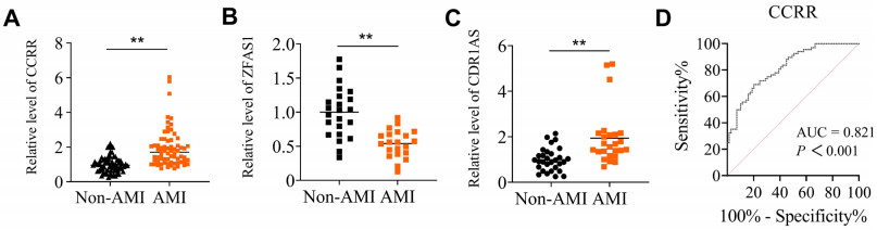
    Zhang Y, Sun L, Xuan L, et al. Reciprocal changes of circulating long non-coding RNAs ZFAS1 and CDR1AS predict acute myocardial infarction. Sci Rep, 2016; 6: 2384.
    [16]
    Wang X, Wang L, Ma Z, et al. Early expressed circulating long noncoding RNA CHAST is associated with cardiac contractile function in patients with acute myocardial infarction. Int J Cardiol, 2020; 302: 15-20. doi: 10.1016/j.ijcard.2019.12.058
    [17]
    Tang Y, Zheng J, Sun Y, et al. MicroRNA-1 regulates cardiomyocyte apoptosis by targeting Bcl-2. Int Heart J, 2009; 50: 377-387. doi: 10.1536/ihj.50.377
    [18]
    Gao L, Liu Y, Guo S, et al. Circulating long noncoding RNA HOTAIR is an essential mediator of acute myocardial infarction. Cell. Physiol. Biochem, 2017; 44: 1497-1508. doi: 10.1159/000485588
    [19]
    Azat M, Huojiahemaiti X, Gao R, et al. Long noncoding RNA MIAT: a potential role in the diagnosis and mediation of acute myocardial infarction. Mol Med Rep, 2019; 20: 5216-5222.
    [20]
    Wang L, Wang L, Wang Q. Constitutive activation of the NEAT1/miR-22-3p/Ltb4r1 signaling pathway in mice with myocardial injury following acute myocardial infarction. Aging (Albany NY), 2021; 13: 15307-15319.
    [21]
    Li M, Wang Y F, Yang X C, et al. Circulating long noncoding RNA LIPCAR acts as a novel biomarker in patients with ST-segment elevation myocardial infarction. Med Sci Monit, 2018; 24: 5064-5070. doi: 10.12659/MSM.909348
    [22]
    Yan L, Zhang Y, Zhang W, et al. lncRNA-NRF is a potential biomarker of heart failure after acute myocardial infarction. J Cardiovasc Transl Res, 2020; 13: 1008-1015. doi: 10.1007/s12265-020-10029-0
    [23]
    Zhang Y, Sun L, Xuan L, et al. Long non-coding RNA CCRR controls cardiac conduction via regulating intercellular coupling. Nat Commun, 2018; 9: 4176. doi: 10.1038/s41467-018-06637-9
    [24]
    Khodayi M, Khalaj-Kondori M, Hoseinpour F M A, et al. Plasma lncRNA profiling identified BC200 and NEAT1 lncRNAs as potential blood-based biomarkers for late-onset Alzheimer's disease. EXCLI J, 2022; 21: 772-785.
    [25]
    Isaac R, Reis F C G, Ying W, et al. Exosomes as mediators of intercellular crosstalk in metabolism. Cell Metab, 2021; 33: 1744-1762. doi: 10.1016/j.cmet.2021.08.006
    [26]
    Zheng M L, Liu X Y, Han R J, et al. Circulating exosomal long noncoding RNAs in patients with acute myocardial infarction. J Cell Mol Med, 2020; 24: 9388-9396. doi: 10.1111/jcmm.15589
    [27]
    Catalano M, O'Driscoll L. Inhibiting extracellular vesicles formation and release: a review of EV inhibitors. J Extracell Vesicles, 2020; 9, 1703244. doi: 10.1080/20013078.2019.1703244
    [28]
    Crea F. How epidemiology can improve the understanding of cardiovascular disease: from mechanisms to treatment. Eur Heart J, 2021; 42: 4503-4507. doi: 10.1093/eurheartj/ehab797
    [29]
    Lerman B, Lerman L O. "Nothing burns like the cold": Cardiovascular disease in frigid zones. Frigid Zone Medicine, 2022; 2 (3): 129-131. doi: 10.2478/fzm-2022-0017
    [30]
    Jin H. Increased risk of cardiovascular disease in cold temperatures. Frigid Zone Medicine, 2022; 2 (3): 138-139. doi: 10.2478/fzm-2022-0020
    [31]
    Oitabén A, Fonseca P, Villanueva M J, et al. Emerging blood-based biomarkers for predicting immunotherapy response in NSCLC. Cancers (Basel), 2022; 14 (11), 2626. doi: 10.3390/cancers14112626
    [32]
    Chaitman B R, Cyr D D, Alexander K P, et al. Cardiovascular and renal implications of myocardial infarction in the ISCHEMIA-CKD trial. Circ Cardiovasc Interv, 2022; 15: e012103.
    [33]
    Wang F, Li X, Xie X, et al. UCA1, a non-protein-coding RNA upregulated in bladder carcinoma and embryo, influencing cell growth and promoting invasion. FEBS Lett, 2008; 582: 1919-1927. doi: 10.1016/j.febslet.2008.05.012
    [34]
    Yan Y, Zhang B, Liu N, et al. Circulating long noncoding RNA UCA1 as a novel biomarker of acute myocardial infarction. Biomed Res Int, 2016; 2016: 8079372.
    [35]
    Zheng P F, Chen L Z, Liu P, et al. A novel lncRNA-miRNA-mRNA triple network identifies lncRNA XIST as a biomarker for acute myocardial infarction. Aging (Albany NY), 2022; 14: 4085-4106.
    [36]
    Wang Y, Zhao R, Liu W, et al. Exosomal circHIPK3 released from hypoxia-pretreated cardiomyocytes regulates oxidative damage in cardiac microvascular endothelial cells via the miR-29a/IGF-1 pathway. Oxid Med Cell Longev, 2019; 2019: 7954657.
    [37]
    Gupta S, Knowlton A A. HSP60 trafficking in adult cardiac myocytes: role of the exosomal pathway. Am J Physiol Heart Circ Physiol, 2007; 292: H3052-3056.
    [38]
    King H W, Michael M Z, Gleadle J M. Hypoxic enhancement of exosome release by breast cancer cells. BMC Cancer, 2021; 12: 421.
    [39]
    Vicencio J M, Yellon D M, Sivaraman V, et al. Plasma exosomes protect the myocardium from ischemia-reperfusion injury. J Am Coll Cardiol, 2015; 65 (15): 1525-1536.
    [40]
    Tomasoni S, Longaretti L, Rota C, et al. Transfer of growth factor receptor mRNA via exosomes unravels the regenerative effect of mesenchymal stem cells. Stem Cells Dev, 2013; 22 (5): 772-778.
    [41]
    Jiao L, Li M, Shao Y, et al. lncRNA-ZFAS1 induces mitochondriamediated apoptosis by causing cytosolic Ca2+ overload in myocardial infarction mice model. Cell Death Dis, 2019; 10: 942.
    [42]
    Shao Y, Li M, Yu Q, et al. CircRNA CDR1as promotes cardiomyocyte apoptosis through activating hippo signaling pathway in diabetic cardiomyopathy. Eur J Pharmacol, 2022; 922: 174915.
  • Relative Articles

    [1]Jiayu Zhu, Jiaqi Zhang, Qi Yu, Liyan Liu, Rennan Feng. Association of dietary phytosterols with prevalence of metabolic dysfunction-associated fatty liver disease in adult population of Northeastern China: An internet-based cross-sectional study[J]. Frigid Zone Medicine, 2025, 5(1): 50-57. doi: 10.1515/fzm-2025-0005
    [2]Jiayue Zhang, Liyao Sun, Xiaohan Yu, Chen Yang, Qi An, Chaoqun Wei, Hongyan Ge. LCN2 aggravates diabetic cataracts by promoting ferroptosis in lens epithelial cells[J]. Frigid Zone Medicine, 2024, 4(3): 177-192. doi: 10.1515/fzm-2024-0018
    [3]Wen Li, Jia Wang, Yilian Yang, Chunlei Duan, Bing Shao, Mingxiu Zhang, Jiapan Wang, Peifeng Li, Ye Yuan, Yan Zhang, Hongyu Ji, Xingda Li, Zhimin Du. Ethanol extract of cassia seed alleviates metabolic dysfunction-associated steatotic liver disease by acting on multiple lipid metabolism-related pathways[J]. Frigid Zone Medicine, 2024, 4(3): 160-176. doi: 10.1515/fzm-2024-0017
    [4]Tongzhu Jin, Zhen Ye, Ruonan Fang, Yue Li, Wei Su, Qianqian Wang, Tianyu Li, Hongli Shan, Yanjie Lu, Haihai Liang. Notum protects against myocardial infarction-induced heart dysfunction by alleviating cardiac fibrosis[J]. Frigid Zone Medicine, 2024, 4(1): 41-50. doi: 10.2478/fzm-2024-0005
    [5]Yanying Wang, Jian Huang, Han Sun, Jie Liu, Yingchun Shao, Manyu Gong, Xuewen Yang, Dongping Liu, Zhuo Wang, Haodong Li, Yanwei Zhang, Xiyang Zhang, Zhiyuan Du, Xiaoping Leng, Lei Jiao, Ying Zhang. Long non-coding RNA-AK138945 regulates myocardial ischemia-reperfusion injury via the miR-1-GRP94 signaling pathway[J]. Frigid Zone Medicine, 2024, 4(1): 31-40. doi: 10.2478/fzm-2024-0004
    [6]Yanan Ni, Dan Liu, Xiaona Zhang, Hong Qiao. Association of point in range with β-cell function and insulin sensitivity of type 2 diabetes mellitus in cold areas[J]. Frigid Zone Medicine, 2023, 3(4): 242-252. doi: 10.2478/fzm-2023-0031
    [7]Manyu Gong, Xuewen Yang, Yaqi Wang, Yanying Wang, Dongping Liu, Haodong Li, Yunmeng Qu, Xiyang Zhang, Yanwei Zhang, Han Sun, Lei Jiao, Ying Zhang. Growth differentiation factor 11 promotes macrophage polarization towards M2 to attenuate myocardial infarction via inhibiting Notch1 signaling pathway[J]. Frigid Zone Medicine, 2023, 3(1): 53-64. doi: 10.2478/fzm-2023-0008
    [8]Zhenghua Li, Kenichi Yamamura. Dissecting pathophysiology of a human dominantly inherited disease, familial amyloidotic polyneuropathy, by using genetically engineered mice[J]. Frigid Zone Medicine, 2022, 2(2): 65-75. doi: 10.2478/fzm-2022-0009
    [9]Yanxia Lin, Huanrui Zhang, Shijie Zhao, Guohui Hua, Wen Tian. The influence of extreme cold ambient temperature on out of hospital cardiac arrest: A systemic review and meta-analysis[J]. Frigid Zone Medicine, 2022, 2(3): 186-192. doi: 10.2478/fzm-2022-0025
    [10]Meijiao He, Yanxiang Zang, Danghui Sun, Jianqiang Li, Guangzhong Liu, Jing Shi, Yue Li. Ticagrelor versus clopidogrel in East Asian patients with acute coronary syndrome: A meta-analysis[J]. Frigid Zone Medicine, 2022, 2(1): 30-40. doi: 10.2478/fzm-2022-0004
    [11]Xiuxiu Wang, Bingjie Hua, Meixi Yu, Shenzhen Liu, Wenya Ma, Fengzhi Ding, Qi Huang, Lai Zhang, Chongwei Bi, Ye Yuan, Mengyu Jin, Tianyi Liu, Ying Yu, Benzhi Cai, Baofeng Yang. Altered expression profile of long non-coding RNAs during heart aging in mice[J]. Frigid Zone Medicine, 2022, 2(2): 109-118. doi: 10.2478/fzm-2022-0015
    [12]Jinglin Wang, Lingfeng Zha. RNA modification by M6A methylation in cardiovascular diseases: Current trends and future directions[J]. Frigid Zone Medicine, 2022, 2(3): 158-177. doi: 10.2478/fzm-2022-0023
    [13]Jinxin Liu, Shaohong Fang, Shanjie Wang, Changbin Sun, Rong Sun, Hengxuan Cai, Bo Yu. Effects of intermittent cold-exposure on culprit plaque morphology in ST-segment elevation myocardial infarction patients: a retrospective study based on optical coherence tomography[J]. Frigid Zone Medicine, 2022, 2(2): 82-89. doi: 10.2478/fzm-2022-0011
    [14]Daba Tolessa Muleta, Xiang Huang, Timur Yagudin, Ying Yang, Jiangang Wang, Xiaoyu Fu, Yue Zhao, Haiyu Gao, Yang Zhou, Zhenwei Pan, Xiangyu Zhong. Ablation of apoptosis-stimulating of p53 protein 1 protects mice from acute hepatic injury and dysfunction via NF-кB pathway in CCl4-induced hepatotoxicity[J]. Frigid Zone Medicine, 2021, 1(1): 53-64. doi: 10.2478/fzm-2021-0007
    [15]Jia Chen, Jinghua Luo, Hao Liu, Xue Du, Shan Zhang, Zizhen Wang, Liu He, Zhichun Feng. Ibuprofen treatment for patent ductus arteriosus in preterm infants: a retrospective cohort study in a leading Chinese center[J]. Frigid Zone Medicine, 2021, 1(2): 103-110. doi: 10.2478/fzm-2021-0013
    [16]Ningning Ma, Jing Chen, Jin Ren. Role of microRNAs in the process of metformin treating multiple diseases[J]. Frigid Zone Medicine, 2021, 1(2): 69-78. doi: 10.2478/fzm-2021-0009
    [17]Yan Feng, Ying Li, Xinling Yang, Limin Han, Luning Wang, Shan Gao, Ruixue Yin, Xue Wang, Jiayang Li, Meiming Liu, Baiyan Li. Direct evidence of VEGF-mediated neuroregulation and afferent explanation of blood pressure dysregulation during angiogenic therapy[J]. Frigid Zone Medicine, 2021, 1(2): 119-126. doi: 10.2478/fzm-2021-0015
    [18]De-an Guo, Wenlong Wei, Changliang Yao, Jianqing Zhang, Qirui Bi. Chinese herbal medicines with beneficial effects[J]. Frigid Zone Medicine, 2021, 1(2): 79-83. doi: 10.2478/fzm-2021-0010
    [19]Xin Xing, Shiqiang Wang. Mammalian hibernation: a unique model for medical research[J]. Frigid Zone Medicine, 2021, 1(2): 65-68. doi: 10.2478/fzm-2021-0008
    [20]Yanyan Liu, Yahan Yu, Xinyao Wang, Guanqun Liu, Xinda Yin, Yunlong Bai, Zhimin Du. Overexpression of microRNA-135b-5p attenuates acute myocardial infarction injury through its antioxidant and anti-apptotic properties[J]. Frigid Zone Medicine, 2021, 1(2): 85-94. doi: 10.2478/fzm-2021-0011
  • 加载中

Catalog

    通讯作者: 陈斌, bchen63@163.com
    • 1. 

      沈阳化工大学材料科学与工程学院 沈阳 110142

    1. 本站搜索
    2. 百度学术搜索
    3. 万方数据库搜索
    4. CNKI搜索

    Figures(7)  / Tables(4)

    Article Metrics

    Article views (179) PDF downloads(7) Cited by()
    Proportional views
    Related

    /

    DownLoad:  Full-Size Img  PowerPoint
    Return
    Return