 投稿系统
投稿系统

Peroxisome proliferator-activated receptors gama ameliorates liver fibrosis in non-alcoholic fatty liver disease by inhibiting TGF-β/Smad signaling activation
doi: 10.2478/fzm-2024-0002
-
Abstract:
Background Nonalcoholic fatty liver disease (NAFLD) is a chronic condition characterized by a progressive decline in liver function, leading to disruptions in liver integrity and metabolic function, resulting in lipid deposition and excessive accumulation of extracellular matrix (ECM). The pathogenesis of NAFLD is complex and not yet fully understood, contributing to the absence of specific therapeutic strategies. Peroxisome proliferator-activated receptor gamma (PPARγ) is a ligand-activated transcription factor pivotal in regulating lipid and glucose metabolism. However, the impacts of PPARγ on NAFLD remains insufficiently explored. Thus, this study aimed to investigate the role of PPARγ in NAFLD and its underlying molecular mechanisms. Methods Chemical detection kits were utilized to quantify collagen content, alanine aminotransferase (ALT), and aspartate aminotransferase (AST) level variations. Quantitative real-time polymerase chain reaction (qRT-PCR) was employed to assess alterations in extracellular matrix-related genes and inflammatory response genes in liver tissue and HepG2 cells, while western blotting was conducted to analyze the levels of both PPARγ and the TGF-β/Smad signaling pathway. Results Our findings unveiled significantly reduced PPARγ expression in a rat model of NAFLD, leading to subsequent activation of the TGF-β/Smad signaling pathway. Furthermore, PPARγ activation effectively mitigated NAFLD progression by inhibiting inflammation and fibrosis-related gene expression and collagen production. On a cellular level, PPARγ activation was found to inhibit the expression of extracellular matrix-related genes such as matrix metalloproteinase 2 (MMP2) and matrix metalloproteinase 9 (MMP9), along with inflammatory response genes interleukin (IL)-1β and IL-6. Additionally, PPARγ activation led to a significant decrease in the levels of ALT and AST. At the molecular level, PPARγ notably down-regulated the TGF-β/Smad signaling pathway, which is known to promote liver fibrosis. Conclusion These groundbreaking findings underscore PPARγ activation as a promising therapeutic approach to delay NAFLD progression by targeting the TGF-β/Smad signaling pathway in hepatic cells. This highlights the potential of PPARγ as a promising therapeutic target for NAFLD management in clinical settings. -
Key words:
- NAFLD /
- PPARγ /
- TGF-β/Smad /
- liver fibrosis
-

Figure 1. Fibrosis and collagen deposition in a rat model of nonalcoholic fatty liver disease (NAFLD). (A) Triglycerides (TG), total cholesterol (TCH), low-density lipoprotein (LDL) and high-density lipoprotein (HDL) concentrations in the control and NAFLD groups. (N = 4-6), *P < 0.05, **P < 0.01, vs. Control. (B) The alanine aminotransferase (ALT) and aspartate aminotransferase (AST) levels in the control and NAFLD groups. (N = 5-6), **P < 0.01, vs. Control. (C) Collagen contents in the liver homogenization samples, indicating collagen deposition in liver tissues in the control and NAFLD groups. (N = 3), **P < 0.01, vs. Control. (D) Oil red O staining of tissue sections showing the lipid deposition in the liver. Scale bar indicates 200 μm. (N = 3). (E) type Ⅰ collagens (Col-1), type Ⅲ collagens (Col-3), matrix metalloproteinase 2 (MMP2), and matrix metalloproteinase 9 (MMP9) mRNA expression in the control and NAFLD groups. (N = 3-11), **P < 0.01, vs. Control. Data are presented as means ± SEM.

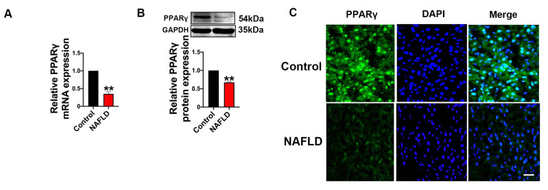
Figure 2. The expression of peroxisome proliferator-activated receptor gamma (PPARγ) was decreased in nonalcoholic fatty liver disease (NAFLD). (A, B) Relative mRNA and protein levels of PPARγ in the control and NAFLD groups. (N = 9), **P < 0.01, vs. Control. (C) Immunofluorescence results indicating the expression of PPARγ in the control and NAFLD groups. Scale bars: 100 μm.

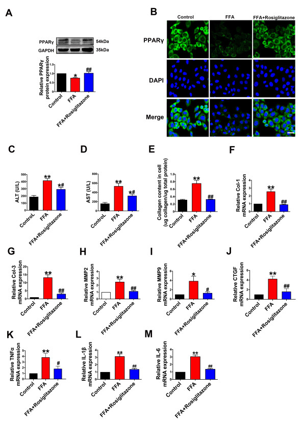
Figure 3. Effect of peroxisome proliferator-activated receptor gamma (PPARγ) inhibition on hepatic fibrosis in nonalcoholic fatty liver disease (NAFLD). (A) Relative protein levels of PPARγ in the control, free fatty acid (FFA), and FFA+Rosiglitazone groups. (N = 6), *P < 0.05, **P < 0.01, vs. Control; ##P < 0.01, vs. FFA. (B) Immunofluorescence results indicating the expression of PPARγ in the control, FFA, and FFA+Rosiglitazone groups. Scale bars: 50 μm. (C, D) ALT and AST concentrations in the control, FFA, and FFA+Rosiglitazone groups. (N = 5), *P < 0.05, **P < 0.01, vs. Control; #P < 0.05, vs. FFA. (E) Collagen contents in the control, FFA, and FFA+Rosiglitazone groups. (N = 5), **P < 0.01, vs. Control; ##P < 0.01, vs. FFA. (F-I) Col-1, Col-3, matrix metalloproteinase 2 (MMP2) and MMP9 mRNA levels in the control, FFA, and FFA+Rosiglitazone groups. (N = 4-5), *P < 0.05, **P < 0.01, vs. Control; #P < 0.05, ##P < 0.01, vs. FFA. (J) The mRNA levels of connective tissue growth factor (CTGF) in the control, FFA, and FFA+Rosiglitazone groups. (N = 4), **P < 0.01, vs. Control; ##P < 0.01, vs. FFA. (K) Tumor necrosis factor-α (TNF-α) mRNA levels in the control, FFA, and FFA + Rosiglitazone groups. (N = 4), **P < 0.01, vs. Control; #P < 0.05, vs. FFA. (L)Interleukin (IL)-1β mRNA level in the control, FFA, and FFA + Rosiglitazone groups. (N = 4), **P < 0.01, vs. Control; ##P < 0.01, vs. FFA. (M) The mRNA levels of IL-6 in the control, FFA, and FFA + Rosiglitazone groups. (N = 4), **P < 0.01, vs. Control; ##P < 0.01, vs. FFA. Data are presented as means ± SEM.

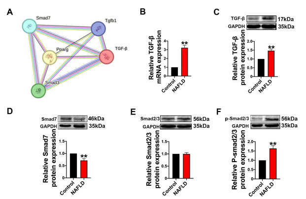
Figure 4. Changes of the transforming growth factor (TGF)-β/Smad signaling pathway in nonalcoholic fatty liver disease (NAFLD). (A) Protein–protein interaction (PPI) network of peroxisome proliferator-activated receptor gamma (PPARγ), TGF-β, Smad2, Smad3, and Smad7 exported from STRING database (
https://string-db.org ) (version 12.0). (B, C) Relative mRNA and protein levels of TGF-β in the control and NAFLD groups. (N = 4-5), **P < 0.01, vs. Control. (D) Relative protein levels of smad7 in each group. (N = 5-8). **P < 0.01, vs. Control. (E) Total smad2/3 protein levels in each group. (N = 3). (F) Phosphorylated protein levels of smad2/3 in each group. (N = 10), **P < 0.01, vs. Control. Data are presented as means ± SEM.
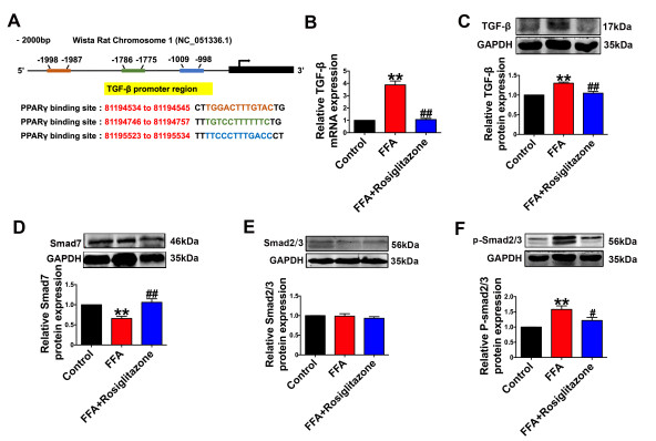
Figure 5. Peroxisome proliferator-activated receptor gamma (PPARγ) inhibits the activation of the transforming growth factor (TGF)-β/Smad axis in nonalcoholic fatty liver disease (NAFLD). (A) PROMO database showing the sequences of the potential binding sites in the proximal region (2000 bp upstream) of TGF-β promoters. (B, C) Relative mRNA and protein levels of TGF-β in the control, free fatty acid (FFA), and FFA+Rosiglitazone groups. (N = 4-5), **P < 0.01, vs. Control; ##P < 0.01, vs. FFA. (D) Relative protein levels of Smad7 in the control, FFA, and FFA+Rosiglitazone groups. (N = 4), **P < 0.01, vs. Control; ##P < 0.01, vs. FFA. (E, F) Relative protein levels Smad2/3 and p-Smad2/3 expression in the control, FFA, and FFA+Rosiglitazone groups. (N = 3-4), **P < 0.01, vs. Control; #P < 0.05, vs. FFA. Data are presented as means ± SEM.

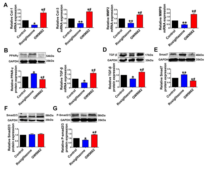
Figure 6. Effect of peroxisome proliferator-activated receptor gamma (PPARγ) on the transforming growth factor (TGF)-β/Smad signaling pathway. (A) The mRNA expression levels of Col-1, Col-3, matrix metalloproteinase 2 (MMP2) and MMP9 in the control, rosiglitazone and GW9662 groups. (N = 3-5), *P < 0.05, **P < 0.01 vs. Control; #P < 0.05, vs. Rosiglitazone. (B) The protein levels of PPARγ in the control, rosiglitazone, and GW9662 groups. (N = 3), *P < 0.05, vs. Control; #P < 0.05, vs. Rosiglitazone. (C, D) Relative TGF-β mRNA and protein levels measured by real-time PCR and western blot analysis, respectively, in the control, rosiglitazone, and GW9662 groups. (N = 4-6), *P < 0.05, vs. Control; #P < 0.05, vs. Rosiglitazone. (E) Relative protein levels of Smad7 in the control, rosiglitazone, and GW9662 groups. (N = 3), *P < 0.05, vs. Control; #P < 0.05, vs. Rosiglitazone. (F) Total smad2/3 protein in the control, rosiglitazone and GW9662 groups. (N = 3). (G) Phosphorylation of smad2/3 in the control, rosiglitazone, and GW9662 groups. (N = 4), *P < 0.05, vs. Control; #P < 0.05, vs. Rosiglitazone. Data are presented as means ± SEM.

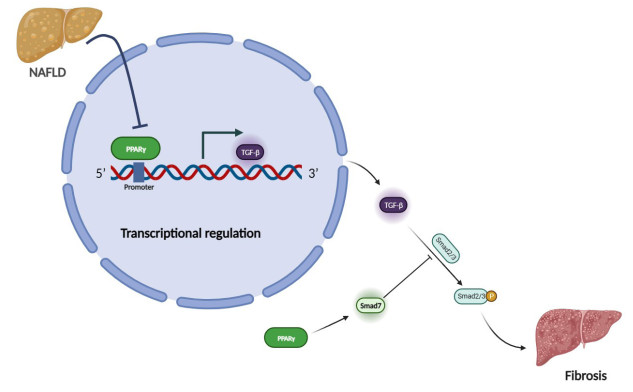
Figure 7. Schematic diagram of the regulation of PPARγ on nonalcoholic fatty liver disease.
Table 1. The sequences of primers for used for qRT-PCR in this study.
Gene Forward primer Reverse primer PPARγ ACCACTCCCATTCCTTTG CACAGACTCGGCACTCG TGF-β GCGAGCGAAGCGACGAGGAG TGGGCGGGATGGCATCAAGGTA Col-1 CAATGGCACGGCTGTGTGCG CACTCGCCCTCCCGTCTTTGG Col-3 TGAATGGTGGTTTTCAGTTCAG GATCCCATCAGCTTCAGAGACT MMP2 AGCTTTGATGGCCCCTATCT GGAGTGACAGGTCCCAGTGT MMP9 CACTGTAACTGGGGGCAACT CACTTCTTGTCAGCGTCGAA IL-1β CCCTGCAGCTGGAGAGTGTGG TGTGCTCTGCTTGAGAGGTGCT IL-6 TTCCTACCCCAACTTCCAATG ATGAGTTGGATGGTCTTGGTC TNFα CCTCTCTCTAATCAGCCCTCTG GAGGACCTGGGAGTAGATGAG CTGF CAGCATGGACGTTCGTCTG AACCACGGTTTGGTCCTTGG Timp1 CTTCTGCAATTCCGACCTCGT ACGCTGGTATAAGGTGGTCTG GAPDH AAGAAGGTGGTGAAGCAGGC TCCACCACCCAGTTGCTGTA PPARγ, peroxisome proliferator-activated receptor gamma; TGF-β, transforming growth factor beta 1; Col-1, collagen, type Ⅰ, alpha 1; Col-3, collagen type Ⅲ alpha 1; MMP2, matrix metallopeptidase 2; MMP9, matrix metallopeptidase 9; IL-1β, interleukin 1 beta; IL-6, interleukin 6; TNFα, tumor necrosis factor-α; CTGF, connective tissue growth factor; Timp1, Tissue inhibitor of metalloproteinases-1; GAPDH, glyceraldehyde-3-phosphate dehydrogenase.  下载: 导出CSV
-
[1] Le M H, Le D M, Baez T C, et al. Global incidence of non-alcoholic fatty liver disease: A systematic review and meta-analysis of 63 studies and 1, 201, 807 persons. J Hepatol, 2023; 79(2): 287-295. doi: 10.1016/j.jhep.2023.03.040 [2] Chen H, Zhan Y, Zhang J, et al. The global, regional, and national burden and trends of NAFLD in 204 Countries and territories: an analysis from global burden of disease 2019. JMIR Public Health Surveill, 2022; 8(12): e34809. doi: 10.2196/34809 [3] Xia M, Sun X, Zheng L, et al. Regional difference in the susceptibility of non-alcoholic fatty liver disease in China. BMJ Open Diabetes Res Care, 2020; 8(1): e001311. doi: 10.1136/bmjdrc-2020-001311 [4] Liu S, Liu Y, Wan B, et al. Association between vitamin D status and non-alcoholic fatty liver disease: a population-based study. J Nutr Sci Vitaminol (Tokyo), 2019; 65: 303-308. doi: 10.3177/jnsv.65.303 [5] Jain R, Wade G, Ong I, et al. Determination of tissue contributions to the circulating lipid pool in cold exposure via systematic assessment of lipid profiles. J Lipid Res, 2022; 63(7): 100197. doi: 10.1016/j.jlr.2022.100197 [6] Su D, Zhou T, Wang Y, et al. Cold exposure regulates hepatic glycogen and lipid metabolism in newborn goats. Int J Mol Sci, 2023; 24(18): 14330. doi: 10.3390/ijms241814330 [7] Heda R, Yazawa M, Shi M, et al. Non-alcoholic fatty liver and chronic kidney disease: Retrospect, introspect, and prospect. World J Gastroenterol, 2021; 27(17): 1864-1882. doi: 10.3748/wjg.v27.i17.1864 [8] Wang S, Tang C, Zhao H, et al. Network pharmacological analysis and experimental validation of the mechanisms of action of Si-Ni-San against liver fibrosis. Front Pharmacol, 2021; 12: 656115. doi: 10.3389/fphar.2021.656115 [9] Wei M, Yan X, Xin X, et al. Hepatocyte-specific Smad4 deficiency alleviates liver fibrosis via the p38/p65 pathway. Int J Mol Sci, 2022; 23(19): 11696. doi: 10.3390/ijms231911696 [10] Li Z, Wang Z, Dong F, et al. Germacrone attenuates hepatic stellate cells activation and liver fibrosis via regulating multiple signaling pathways. Front Pharmacol, 2021; 12: 745561. doi: 10.3389/fphar.2021.745561 [11] Yuan S, Wei C, Liu G, et al. Sorafenib attenuates liver fibrosis by triggering hepatic stellate cell ferroptosis via HIF-1alpha/SLC7A11 pathway. Cell Prolif, 2022; 55(1): e13158. doi: 10.1111/cpr.13158 [12] Seo H Y, Lee S H, Han E, et al. Evogliptin directly inhibits inflammatory and fibrotic signaling in isolated liver cells. Int J Mol Sci, 2022; 23(19): 11636. doi: 10.3390/ijms231911636 [13] Song Y, Wei J, Li R, et al. Tyrosine kinase receptor B attenuates liver fibrosis by inhibiting TGF-beta/SMAD signaling. Hepatology, 2023; 78(5): 1433-1447. doi: 10.1097/HEP.0000000000000319 [14] Vallee A, Vallee J N, Lecarpentier Y. PPARgamma agonists: potential treatment for autism spectrum disorder by inhibiting the canonical WNT/beta-catenin pathway. Mol Psychiatry, 2019; 24(5): 643-652. doi: 10.1038/s41380-018-0131-4 [15] Khan R S, Bril F, Cusi K, et al. Modulation of insulin resistance in nonalcoholic fatty liver disease. Hepatology, 2019; 70(2): 711-724. doi: 10.1002/hep.30429 [16] Trauner M, Fuchs C D. Novel therapeutic targets for cholestatic and fatty liver disease. Gut, 2022; 71(1): 194-209. doi: 10.1136/gutjnl-2021-324305 [17] Snyder H S, Sakaan S A, March K L, et al. Non-alcoholic fatty liver disease: a review of anti-diabetic pharmacologic therapies. J Clin Transl Hepatol, 2018; 6(2): 168-174. doi: 10.14218/JCTH.2017.00050 [18] Chang E, Park C Y, Park S W. Role of thiazolidinediones, insulin sensitizers, in non-alcoholic fatty liver disease. J Diabetes Investig, 2013; 4(6): 517-524. doi: 10.1111/jdi.12107 [19] Li J, Gong L, Zhang R, et al. Fibroblast growth factor 21 inhibited inflammation and fibrosis after myocardial infarction via EGR1. Eur J Pharmacol, 2021; 910: 174470. doi: 10.1016/j.ejphar.2021.174470 [20] Zhang Q, Chen X, Hu Y, et al. BIRC5 inhibition is associated with pyroptotic cell death via Caspase3-GSDME pathway in lung adenocarcinoma cells. Int J Mol Sci, 2023; 24(19): 14663. doi: 10.3390/ijms241914663 [21] Li J, Li Y, Liu Y, et al. Fibroblast growth factor 21 ameliorates Na(V)1.5 and Kir2.1 channel dysregulation in human AC16 cardiomyocytes. Front Pharmacol, 2021; 12: 715466. doi: 10.3389/fphar.2021.715466 [22] Li J, Xu C, Liu Y, et al. Fibroblast growth factor 21 inhibited ischemic arrhythmias via targeting miR-143/EGR1 axis. Basic Res Cardiol, 2020; 115(2): 9. doi: 10.1007/s00395-019-0768-4 [23] Zhou Q Y, Yang H M, Liu J X, et al. MicroRNA-497 induced by Clonorchis sinensis enhances the TGF-beta/Smad signaling pathway to promote hepatic fibrosis by targeting Smad7. Parasit Vectors, 2021; 14(1): 472. doi: 10.1186/s13071-021-04972-3 [24] Ganai A A, Husain M. Genistein attenuates D-GalN induced liver fibrosis/chronic liver damage in rats by blocking the TGF-beta/Smad signaling pathways. Chem Biol Interact, 2017; 261: 80-85. doi: 10.1016/j.cbi.2016.11.022 [25] Porcuna J, Minguez-Martinez J and Ricote M. The PPARalpha and PPARgamma epigenetic landscape in cancer and immune and metabolic disorders. Int J Mol Sci, 2021; 22(19): 10573. doi: 10.3390/ijms221910573 [26] Hendawy O, Gomaa H A M, Hussein S, et al. Cold-pressed raspberry seeds oil ameliorates high-fat diet triggered non-alcoholic fatty liver disease. Saudi Pharm J, 2021; 29(11): 1303-1313. doi: 10.1016/j.jsps.2021.09.014 [27] Wu X, Cheng B, Guo X, et al. PPARalpha/gamma signaling pathways are involved in Chlamydia pneumoniae-induced foam cell formation via upregulation of SR-A1 and ACAT1 and downregulation of ABCA1/G1. Microb Pathog, 2021; 161(Pt B): 105284. doi: 10.1016/j.micpath.2021.105284 [28] Shan S, Zhou J, Yin R, et al. Millet bran protein hydrolysate displays the anti-non-alcoholic fatty liver disease effect via activating peroxisome proliferator-activated receptor gamma to restrain fatty acid uptake. J Agric Food Chem, 2023; 71(3): 1628-1642. doi: 10.1021/acs.jafc.2c08169 [29] He J, Hong B, Bian M, et al. Docosahexaenoic acid inhibits hepatic stellate cell activation to attenuate liver fibrosis in a PPARgamma-dependent manner. Int Immunopharmacol, 2019; 75: 105816. doi: 10.1016/j.intimp.2019.105816 [30] Fiorucci S, Distrutti E. Linking liver metabolic and vascular disease via bile acid signaling. Trends Mol Med, 2022; 28(1): 51-66. doi: 10.1016/j.molmed.2021.10.005 [31] Kim E R, Park J S, Kim J H, et al. A GLP-1/GLP-2 receptor dual agonist to treat NASH: targeting the gut-liver axis and microbiome. Hepatology, 2022; 75(6): 1523-1538. doi: 10.1002/hep.32235 [32] Brougham-Cook A, Jain I, Kukla D A, et al. High throughput interrogation of human liver stellate cells reveals microenvironmental regulation of phenotype. Acta Biomater, 2022; 138: 240-253. doi: 10.1016/j.actbio.2021.11.015 [33] Wu G, Liu Y, Feng W, et al. Hypoxia-induced adipose lipolysis requires fibroblast growth factor 21. Front Pharmacol, 2020; 11: 1279. doi: 10.3389/fphar.2020.01279 [34] Chojkier M, Lyche K D, Filip M. Increased production of collagen in vivo by hepatocytes and nonparenchymal cells in rats with carbon tetrachloride-induced hepatic fibrosis. Hepatology, 1988; 8(4): 808-814. doi: 10.1002/hep.1840080419 [35] Wehrhan F, Hyckel P, Guentsch A, et al. Bisphosphonate-associated osteonecrosis of the jaw is linked to suppressed TGFbeta1-signaling and increased Galectin-3 expression: a histological study on biopsies. J Transl Med, 2011; 9: 102. doi: 10.1186/1479-5876-9-102 [36] Hilt Z T, Maurya P, Tesoro L, et al. Beta2M signals monocytes through non-canonical TGFbeta receptor signal transduction. Circ Res, 2021; 128(5): 655-669. doi: 10.1161/CIRCRESAHA.120.317119 [37] Zhong C, Lin Z, Ke L, et al. Recent research progress (2015-2021) and perspectives on the pharmacological effects and mechanisms of tanshinone IIA. Front Pharmacol, 2021; 12: 778847. doi: 10.3389/fphar.2021.778847 [38] Tian H, Liu H, Yu J, et al. PHF14 enhances DNA methylation of SMAD7 gene to promote TGF-beta-driven lung adenocarcinoma metastasis. Cell Discov, 2023; 9(1): 41. doi: 10.1038/s41421-023-00528-0 -
 fzm-4-1-12_ESM.pdf

-

 点击查看大图

计量
- 文章访问数: 1154
- HTML全文浏览量: 625
- PDF下载量: 32
- 被引次数: 0

