 投稿系统
投稿系统

Hydrogen sulfide, microbiota, and sulfur amino acid restriction diet
doi: 10.2478/fzm-2021-0003
-
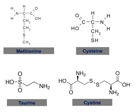
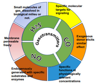
Abstract: Eukaryotes and microbiota produce H2S, using the same substrates and enzymes which constitute the reverse-trans-sulfuration and transsulfuration pathways. The homeostasis of gut microbiota impacts on the structural and functional integrity of gut epithelial barrier. Microbiota also serve as signalling sources to inform the host of the metabolism and functional changes. Microbiota dysbiosis negatively affect human health, contributing to diseases like obesity, diabetes, inflammatory bowel diseases, and asthma. Not by coincidence, these pathological conditions are also closely related to the abnormal metabolism and function of H2S signalling.H2S serves as a bacterial signal to the host and the host-produced H2S impacts on the population and size of microbiota. These bi-directional interactions become especially important for the digestion and utilization of sulfur amino acid in diet. Dietary restriction of sulfur amino acid increases the endogenous production of H2S by the host and consequently offers many health benefits. It, on the other hand, decreases the nutritional supply to the microbiota, which could be remedied by the co-application of prebiotics and probiotics. It is strategically sound to target the expression of H2S-producing enzymes in different organs to slow aging processes in our body and promote better health.
-
[1] Wang R. Two's company, three's a crowd: Can H2S be the third endogenous gaseous transmitter? FASEB J, 2002; 16: 1792-1798. doi: 10.1096/fj.02-0211hyp [2] Wang R. Physiological implications of hydrogen sulfide: a whiff exploration that blossomed. Physiol Rev, 2012; 92(2): 791-896. doi: 10.1152/physrev.00017.2011 [3] Wang R. Gasotransmitters: growing pains and joys. Trends Biochem Sci, 2014; 39(5): 60-72. http://blog.sciencenet.cn/home.php/blog.sciencenet.cn/home.php?mod=attachment&filename=Gasotransmitters%20growing%20pains%20and%20joys.pdf&id=53483 [4] Wang R. Toxic gas, lifesaver. Sci American, 2010; 302(3): 66-71. doi: 10.1038/scientificamerican0310-66 [5] Cavallini D, De Marco C, Mondovi B, et al. The cleavage of cystine by cystathionase and the transulfuration of hypotaurine. Enzymologia, 1960; 22: 161-173. http://europepmc.org/abstract/MED/13691729 [6] Selim A S, Greenberg D M. An enzyme that synthesizes cystathionine and deaminates L-serine. J Biol Chem, 1959; 234(6): 1474-1480. doi: 10.1016/S0021-9258(18)70033-1 [7] Kun E, Fanshier D W. Inhibition of beta-mercaptopyruvate transsulfurase by metal chelate compounds. Biochim Biophys Acta, 1961; 48: 187-188. doi: 10.1016/0006-3002(61)90531-5 [8] Erickson P F, Maxwell I H, Su L J, et al. Sequence of cDNA for rat cystathionine gamma-lyase and comparison of deduced amino acid sequence with related Escherichia coli enzymes. Biochem J, 1990; 269(2): 335-340. doi: 10.1042/bj2690335 [9] Kraus J P, Williamson C L, Firgaira F A, et al. Cloning and screening with nanogram amounts of immunopurified mRNAs: cDNA cloning and chromosomal mapping of cystathionine beta-synthase and the beta subunit of propionyl-CoA carboxylase. Proc Natl Acad Sci USA, 1986; 83(7): 2047-2051. doi: 10.1073/pnas.83.7.2047 [10] Pallini R, Guazzi G C, Cannella C, et al. Cloning and sequence analysis of the human liver rhodanese: comparison with the bovine and chicken enzymes. Biochem Biophys Res Commun, 1991; 180(2): 887-893. doi: 10.1016/S0006-291X(05)81148-9 [11] Zhao W, Zhang J, Lu Y, et al. The vasorelaxant effect of H2S as a novel endogenous gaseous KATP channel opener. EMBO J, 2001; 20: 6008-6016. doi: 10.1093/emboj/20.21.6008 [12] Stipanuk M H, Beck P W. Characterization of the enzymic capacity for cysteine desulphhydration in liver and kidney of the rat. Biochem J, 1982; 206(2): 267-277. doi: 10.1042/bj2060267 [13] Goodwin L R, Francom D, Dieken F P, et al. Determination of sulfide in brain tissue by gas dialysis/ion chromatography: Postmortem studies and two case reports. J Anal Toxicol, 1989; 13(2): 105-109. doi: 10.1093/jat/13.2.105 [14] Savage J C, Gould D H. Determination of sulfide in brain tissue and rumen fluid by ion-interaction reversed-phase high-performance liquid chromatography. J Chromatogr, 1990; 526(2): 540-545. http://pdf.eurekamag.com/001/001795932.pdf [15] Fu M, Zhang W, Wu L, et al. Hydrogen sulfide (H2S) metabolism in mitochondria and its regulatory role in energy production. Proc Natl Acad Sci USA, 2012; 109(8): 2943-2948. doi: 10.1073/pnas.1115634109 [16] Módis K, Ju Y, Ahmad A, et al. S-sulfhydration of ATP synthase by hydrogen sulfide stimulates mitochondrial bioenergetics. Pharmacol Res, 2016; 113(Pt A): 116-124. http://www.ncbi.nlm.nih.gov/pmc/articles/PMC5107138/pdf/nihms812695.pdf [17] Abe K, Kimura H. The possible role of hydrogen sulfide as an endogenous neuromodulator. J Neurosci, 1996; 16(3): 1066-1071. doi: 10.1523/JNEUROSCI.16-03-01066.1996 [18] Hosoki R, Matsuki N, Kimura H. The possible role of hydrogen sulfide as an endogenous smooth muscle relaxant in synergy with nitric oxide. Biochem Biophys Res Commun, 1997; 237: 527-531. doi: 10.1006/bbrc.1997.6878 [19] Yang G, Wu L, Jiang B, et al. H2S as a physiologic vasorelaxant: hypertension in mice with deletion of cystathionine gamma-lyase. Science, 2008; 322: 587-590. doi: 10.1126/science.1162667 [20] Mani S, Li H, Untereiner A, et al. Decreased endogenous production of hydrogen sulfide accelerates atherosclerosis. Circulation, 2013; 127(25): 2523-2534. doi: 10.1161/CIRCULATIONAHA.113.002208 [21] Yang G, Wu L, Bryan S, et al. Cystathionine gamma-lyase deficiency and overproliferation of smooth muscle cells. Cardiovasc Res, 2010; 86(3): 487-495. doi: 10.1093/cvr/cvp420 [22] Altaany Z, Ju Y, Yang G, et al. The coordination of S-sulfhydration, S-nitrosylation, and phosphorylation of endothelial nitric oxide synthase by hydrogen sulfide. Sci Signal, 2014; 7 (342): ra87. http://www.onacademic.com/detail/journal_1000037961475910_2f53.html [23] Papapetropoulos A, Pyriochou A, Altaany Z, et al. Hydrogen sulfide is an endogenous stimulator of angiogenesis. Proc Natl Acad Sci USA, 2009; 106(51): 21972-21977. doi: 10.1073/pnas.0908047106 [24] Jiang B, Tang G, Cao K, et al. Molecular mechanism for H2S-induced activation of KATP channels. Antioxid Redox Signal, 2010; 12(10): 1167-1178. doi: 10.1089/ars.2009.2894 [25] Tang G, Wu L, Liang W, et al. Direct stimulation of KATP channels by exogenous and endogenous hydrogen sulfide in vascular smooth muscle cells. Mol Pharmacol, 2005; 68(6): 1757-1764. doi: 10.1124/mol.105.017467 [26] Tang G, Yang G, Jiang B, et al. H2S is an endothelium-derived hyperpolarizing factor. Antioxid Redox Signal, 2013; 19(14): 1634-1646. doi: 10.1089/ars.2012.4805 [27] Mustafa A, Sikka G, Gazi S K, et al. Hydrogen sulfide as endothelium-derived hyperpolarizing factor sulfhydrates potassium channels. Circ Res, 2011; 109(11): 1259-1268. doi: 10.1161/CIRCRESAHA.111.240242 [28] Elrod J W, Calvert J W, Morrison J, et al. Hydrogen sulfide attenuates myocardial ischemia-reperfusion injury by preservation of mitochondrial function. Proc Natl Acad Sci USA, 2007; 104(39): 15560-15565. doi: 10.1073/pnas.0705891104 [29] Johansen D, Ytrehus K, Baxter G F, et al. Exogenous hydrogen sulfide (H2S) protects against regional myocardial ischemia-reperfusion injury -Evidence for a role of KATP channels. Basic Res Cardiol, 2006; 101(1): 53-60. doi: 10.1007/s00395-005-0569-9 [30] Kondo K, Bhushan S, King A L, et al. H2S protects against pressure overload induced heart failure via upregulation of endothelial nitric oxide synthase (eNOS). Circulation, 2013; 127(10): 1116-1127. doi: 10.1161/CIRCULATIONAHA.112.000855 [31] Madden J A, Ahlf S B, Dantuma M W, et al. Precursors and inhibitors of hydrogen sulfide synthesis affect acute hypoxic pulmonary vasoconstriction in the intact lung. J Appl Physiol, 2012; 112(3): 411-418. doi: 10.1152/japplphysiol.01049.2011 [32] Wu L, Yang W, Jia X, et al. Pancreatic islet overproduction of H2S and suppressed insulin release in Zucker diabetic rats. Lab Invest, 2009; 89(1): 59-67. doi: 10.1038/labinvest.2008.109 [33] Vachek H, Wood J L. Purification and properties of mercaptopyruvate sulfur transferase of Escherichia coli. Biochim Biophys Acta, 1972; 258(1): 133-146. doi: 10.1016/0005-2744(72)90973-4 [34] Taniguchi T, Kimura T. Role of 3-mercaptopyruvate sulfurtransferase in the formation of the iron-sulfur chromophore of adrenal ferredoxin. Biochim Biophys Acta, 1974; 364(2): 284-295. doi: 10.1016/0005-2744(74)90014-X [35] Cao X, Ding L, Xie Z Z, et al. A Review of hydrogen sulfide synthesis, metabolism, and measurement: Is modulation of hydrogen sulfide a novel therapeutic for cancer? Antiox Redox Signal, 2019; 31(1): 1-38. doi: 10.1089/ars.2017.7058 [36] Zhao W, Ndisang J F, Wang R. Modulation of endogenous production of H2S in rat tissues. Can J Physiol Pharmacol, 2003; 81(9): 848-853 doi: 10.1139/y03-077 [37] Paul B D, Sbodio J I, Xu R, et al. Cystathionine γ-lyase deficiency mediates neurodegeneration in Huntington's disease. Nature, 2014; 509(7498): 96-100. doi: 10.1038/nature13136 [38] Shibuya N, Mikami Y, Kimura Y, et al. Vascular endothelium expresses 3-mercaptopyruvate sulfurtransferase and produces hydrogen sulfide. J Biochem, 2009; 146(5): 623-626. doi: 10.1093/jb/mvp111 [39] Tomita M, Nagahara N, Ito T. Expression of 3-mercaptopyruvate sulfurtransferase in the mouse. Molecules, 2016; 21(12): 1707. doi: 10.3390/molecules21121707 [40] Vitvitsky V, Yadav P K, Kurthen A, et al. Sulfide oxidation by a noncanonical pathway in red blood cells generates thiosulfate and polysulfides. J Biol Chem, 2015; 290(13): 8310-8320. doi: 10.1074/jbc.M115.639831 [41] Ma N, Ma X. Dietary amino acids and the gut-microbiome-immune axis: Physiological metabolism and therapeutic prospects. Comprehensive Reviews in Food Science and Food Safety, 2019; 18: 221-242. doi: 10.1111/1541-4337.12401 [42] Shen X, Carlstrom M, Borniquel S, et al. Microbial regulation of host hydrogen sulfide bioavailability and metabolism. Free Radic Biol Med, 2013; 60: 195-200. doi: 10.1016/j.freeradbiomed.2013.02.024 [43] Sawa T, Ono K, Tsutsuki H, et al. Reactive cysteine persulphides: occurrence, biosynthesis, antioxidant activity, methodologies, and bacterial persulphide signalling. Adv Microb Physiol, 2018; 72: 1-28. [44] Hine C, Zhu Y, Hollenberg A N, et al. Dietary and endocrine regulation of endogenous hydrogen sulfide production: Implications for longevity. Antioxid Redox Signal, 2018; 28(16): 1483-1502. doi: 10.1089/ars.2017.7434 [45] Shatalin K, Shatalina E, Mironov A, et al. H2S: a universal defense against antibiotics in bacteria. Science, 2011; 334(6058): 986-990. doi: 10.1126/science.1209855 [46] Longchamp A, Mirabella T, Arduini A, et al. Amino acid restriction triggers angiogenesis via GCN2/ATF4 regulation of VEGF and H2S production. Cell, 2018; 173: 117-129. doi: 10.1016/j.cell.2018.03.001 [47] Hine C, Harputlugil E, Zhang Y, et al. Endogenous hydrogen sulfide production is essential for dietary restriction benefits. Cell, 2015; 160(1-2): 132-144. doi: 10.1016/j.cell.2014.11.048 [48] Dong Z, Gao X, Chinchilli V M, et al. Association of sulfur amino acid consumption with cardiometabolic risk factors: Cross-sectional findings from NHANES Ⅲ. E Clinical Medicine, 2020; 19: 100248. http://www.sciencedirect.com/science/article/pii/S2589537019302573 [49] Wang R. The surprising reason eating less meat is linked to a longer life: A smelly toxic gas. Conversation. https://theconversation.com/the-surprising-reason-eating-less-meat-is-linked-to-a-longer-life-a-smelly-toxic-gas-151187. Jan. 19 2021 Accessed. [50] Kabil O, Vitvitsky V, Banerjee R. Sulfur as a signaling nutrient through hydrogen sulfide. Annu Rev Nutr, 2014; 34: 171-205. doi: 10.1146/annurev-nutr-071813-105654 [51] McIsaac R S, Lewis K N, Gibney P A, et al. From yeast to human: exploring the comparative biology of methionine restriction in extending eukaryotic life span. Ann NY Acad Sci, 2016; 1363: 155-170. doi: 10.1111/nyas.13032 [52] Elshorbagy A K, Valdivia-Garcia M, Mattocks D A, et al. Cysteine supplementation reverses methionine restriction effects on rat adiposity: significance of stearoyl-coenzyme A desaturase. J Lipid Res, 2011; 52: 104-112. doi: 10.1194/jlr.M010215 [53] Koziel R, Ruckenstuhl C, Albertini E, et al. Methionine restriction slows down senescence in human diploid fibroblasts. Aging Cell, 2014; 13: 1038-1048. doi: 10.1111/acel.12266 [54] Huxtable R J. Physiological actions of taurine. Physiol Rev, 1992; 72: 101-163. doi: 10.1152/physrev.1992.72.1.101 [55] Kanakubo K, Fascetti A J, Larsen J A. Assessment of protein and amino acid concentrations and labeling adequacy of commercial vegetarian diets formulated for dogs and cats. J Am Veterin Med Assoc, 2015; 247(4): 385-392. doi: 10.2460/javma.247.4.385 [56] Jonsson W O, Margolies N S, Anthony T G. Dietary sulfur amino acid restriction and the integrated stress response: Mechanistic insights. Nutrients, 2019; 11: 1349. doi: 10.3390/nu11061349 [57] Pettit A P, Jonsson W O, Bargoud A R, et al. Dietary methionine restriction regulates liver protein synthesis and gene expression independently of eukaryotic initiation factor 2 phosphorylation in mice. J Nutr, 2017; 147(6): 1031-1040. doi: 10.3945/jn.116.246710 [58] Yadav V, Gao X H, Willard B, et al. Hydrogen sulfide modulates eukaryotic translation initiation factor 2 (eIF2) phosphorylation status in the integrated stress-response pathway. J Biol Chem, 2017; 292: 13143-13153. doi: 10.1074/jbc.M117.778654 [59] Miousse I R, Pathak R, Garg S, et al. Short-term dietary methionine supplementation affects one-carbon metabolism and DNA methylation in the mouse gut and leads to altered microbiome profiles, barrier function, gene expression and histomorphology. Genes Nutr, 2017; 12: 22. doi: 10.1186/s12263-017-0576-0 [60] Hill C, Guarner F, Reid G, et al. The International Scientific Association for Probiotics and Prebiotics consensus statement on the scope and appropriate use of the term probiotic. Nat Rev Gastroenterol Hepatol, 2014; 11: 506-514. doi: 10.1038/nrgastro.2014.66 [61] Abatenh E, Gizaw B, Tsegay Z, et al. Health benefits of probiotics. J Bacteriol Infec Dis, 2018; 2(1): 17-27. [62] Gibson G R, Probert H M, Loo J V, et al. Dietary modulation of the human colonic microbiota: updating the concept of prebiotics. Nutr Res Rev, 2004; 17(2): 259-275. doi: 10.1079/NRR200479 [63] Dai Z, Wu Z, Hang S, et al. Amino acid metabolism in intestinal bacteria and its potential implications for mammalian reproduction. Mol Hum Reprod, 2015; 21(5): 389-409. doi: 10.1093/molehr/gav003 [64] Talbott S, Talbott J. Effect of beta 1, 3/1, 6 glucan on respiratory tract infection symptoms and mood state in marathon athletes. J Sports Sci Med, 2009; 8(4): 509-515. http://mypeacebeverage.com/wp-content/uploads/2014/11/6.-Talbott-California-Marathon-published-Journal-of-Sports-Sci-Med-122009.pdf [65] Savignac H M, Corona G, Mills H, et al. Prebiotic feeding elevates central brain derived neurotrophic factor, N-methyl-D-aspartate receptor subunits and D-serine. Neurochem Int, 2013; 63(8): 756-764. doi: 10.1016/j.neuint.2013.10.006 [66] Yang Z, Liao S F. Physiological effects of dietary amino acids on gut health and functions of swine. Front Vet Sci, 2019; 6: 169. doi: 10.3389/fvets.2019.00169 [67] Matsumoto K, Ichimura M, Tsuneyama K, et al. Fructo-oligosaccharides and intestinal barrier function in a methionine±choline-deficient mouse model of nonalcoholic steatohepatitis. PLoS ONE, 2017; 12(6): e0175406. doi: 10.1371/journal.pone.0175406 [68] Liu X, Cao S, Zhang X. Modulation of Gut Microbiota-Brain Axis by Probiotics, Prebiotics, and diet. J Agric Food Chem, 2015; 63: 7885-7895. doi: 10.1021/acs.jafc.5b02404 [69] Kuhnlein H V, Receveur O, Soueida R, et al. Arctic indigenous peoples experience the nutrition transition with changing dietary patterns and obesity. J Nutr, 2004; 134: 1447-1453. doi: 10.1093/jn/134.6.1447 [70] Lucas M, Dewailly E, Blanchet C, et al. Plasma omega-3 and psychological distress among Nunavik Inuit (Canada). Psychiatry Res, 2009; 167(3): 266-278. doi: 10.1016/j.psychres.2008.04.012 [71] McLaughlin J, Middaugh J, Boudreau D, et al. Adipose tissue triglyceride fatty acids and atherosclerosis in Alaska Natives and non-Natives. Atherosclerosis, 2005; 181(2): 353-362. doi: 10.1016/j.atherosclerosis.2005.01.019 [72] Hu X F, Kenny T A, Chan H M. Inuit country food diet pattern is associated with lower risk of coronary heart disease. J Acad Nutr Diet, 2018; 118(7): 1237-1248. doi: 10.1016/j.jand.2018.02.004 [73] Dordević D, Jančíková S, Vítězová M, Kushkevych I. Hydrogen sulfide toxicity in the gut environment: Meta-analysis of sulfate-reducing and lactic acid bacteria in inflammatory processes. J Adv Res, 2021; 27: 55-69. doi: 10.1016/j.jare.2020.03.003 -

 下载:
下载:

 点击查看大图
图(3)
计量
- 文章访问数: 689
- HTML全文浏览量: 387
- PDF下载量: 4
- 被引次数: 0

