 投稿系统
投稿系统

Silencing lipocalin-2 ameliorates severe hypothermia-induced acute lung injury in rats by regulating macrophage polarization
doi: 10.1515/fzm-2026-0003
-
Abstract:
Objective Severe hypothermia can precipitate acute lung injury (ALI) and may progress to acute respiratory distress syndrome (ARDS). Alveolar macrophage (AM) polarization plays a pivotal role in both the pathogenesis and resolution of ALI/ARDS. This study aimed to investigate alterations in AM polarization and to identify potential regulatory targets of macrophage polarization in severe hypothermia-induced ALI (SH-ALI). Methods An SH-ALI rat model was established by immersion in 15 ℃ seawater for 5 h. Survival rate, inflammatory cytokine levels, lung histopathology, lung wet-to-dry weight ratio, and AM polarization status were assessed. RNA sequencing was performed to identify differentially expressed genes in bronchoalveolar lavage fluid-stimulated AMs from SH-ALI rats compared with normal AMs. Lipocalin-2 (LCN2) was identified and validated as a differentially expressed gene in SH-ALI. The effects of LCN2 on macrophage polarization, survival rate, lung injury severity, and inflammatory factor levels were further evaluated. Results SH-ALI was characterized by increased mortality, elevated inflammatory cytokine levels, marked inflammatory cell infiltration, aggravated pulmonary edema, and significant pulmonary parenchyma injury. An imbalance between M1 and M2 macrophage polarization was observed, with severe hypothermia promoting M1 polarization. LCN2 expression was significantly upregulated in SH-ALI rats. Downregulation of LCN2 suppressed M1 polarization and alleviated lung injury in SH-ALI in rats. Conclusion Immersion in 15 ℃ seawater for 5 h successfully establishes an SH-ALI rat model. SH-ALI is associated with excessive M1 polarization, and LCN2 knockdown mitigates lung injury by inhibiting M1 polarization in SH-ALI rats. -
Key words:
- hypothermia /
- acute lung injury /
- lipocalin-2
-

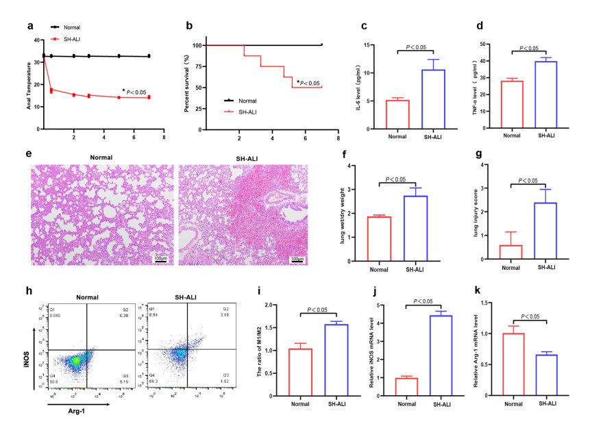
Figure 1. Establishment of the SH-ALI rat model and AM polarization in SH-ALI rats
(a) Rectal temperature of rats in normal and SH-ALI groups. Data are expressed as means ± SD (n = 5). *P < 0.05 vs. normal group. (b) Survival rate of rats in the normal and SH-ALI groups. Survival was monitored for 7 h, and survival curves were compared using the log-rank test. Data are expressed as survival percentage (n = 15). *P < 0.05 vs. normal group. (c-d) Serum levels of inflammatory cytokines IL-6 (c) and TNF-α (d). (e) Representative hematoxylin and eosin (H&E)-stained lung tissue sections. (f) Lung wet-to-dry (W/D) weight ratios in the normal and SH-ALI groups. (g) Lung injury scores in the normal and SH-ALI groups. (h-k) Severe hypothermia promotes M1 polarization: (h) Representative flow cytometry plots showing iNOS and Arg-1 expression in AMs isolated from BALF in each group. (i) Quantitative analysis of the iNOS+/Arg-1+ ratio in each group. (j-k) mRNA expression levels of the M1 marker iNOS (j) and M2 marker Arg1 (k) in AMs isolated from BALF, as determined by qRT-PCR. Data are expressed as means ± SD (n = 5). *P < 0.05 vs. normal group. Abbreviations: SH-ALI, severe hypothermia-induced acute lung injury; AM, alveolar macrophage; IL-6, interleukin-6; TNF-α, tumor necrosis factor-α; H&E, hematoxylin and eosin; BALF, bronchoalveolar lavage fluid; iNOS, inducible nitric oxide synthase; Arg-1, arginase-1.

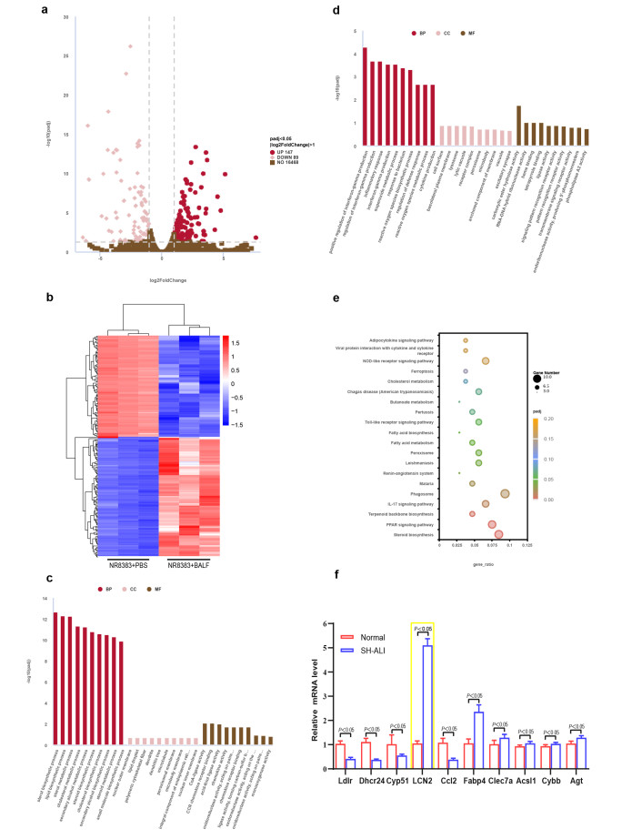
Figure 2. RNA-Seq analysis of NR8383 cells stimulated with BALF from SH-ALI rat
(a) Volcano plot of DEGs between the NR8383+PBS and NR8383+BALF groups. (b) Hierarchical clustering heatmap of DEGs. (c-d) GO enrichment analysis of upregulated (c) and downregulated (d) genes. (e) KEGG pathway enrichment analysis of DEGs. (f) mRNA expression levels of selected differentially expressed genes in AMs isolated from BALF in each group, as determined by qRT-PCR. Data are expressed as means ± SD (n = 3). Abbreviations: PBS, phosphate-buffered saline; BALF, bronchoalveolar lavage fluid; GO, Gene Ontology; KEGG, Kyoto Encyclopedia of Genes and Genomes; AM, alveolar macrophage.

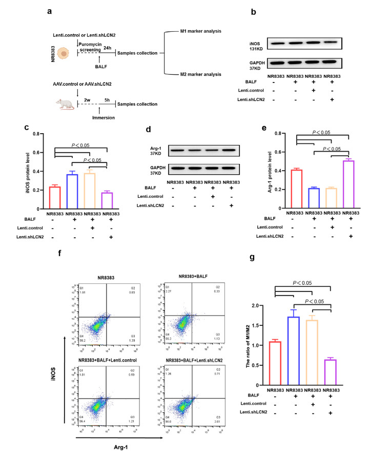
Figure 3. LCN2 knockdown suppresses M1 polarization in NR8383 cells
(a) Schematic diagram of the experimental design showing the time points of viral transfection, seawater immersion, and sample collection. (b) Representative western blot images showing iNOS protein expression (normalized to GAPDH) in each group. (c) Quantitative analysis of iNOS protein expression normalized to GAPDH. (d) Representative western blot images showing Arg-1 protein expression (normalized to GAPDH) in each group. (e) Quantitative analysis of Arg-1 protein expression normalized to GAPDH. (f) Representative flow cytometry plots showing iNOS and Arg-1 expression in each group. (g) Quantitative analysis of the iNOS+/Arg-1+ ratio in each group. Data are expressed as means ± SD (n = 3). Abbreviations: LCN2, lipocalin-2; iNOS, inducible nitric oxide synthase; GAPDH, glyceraldehyde-3-phosphate dehydrogenase; Arg-1, arginase-1.

Figure 4. LCN2 knockdown suppresses M1 polarization in SH-ALI rats
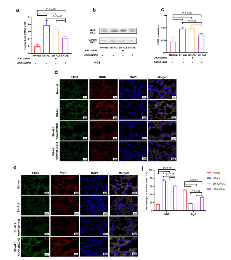
(a) RelativLCN2 mRNA levels normalized to GAPDH in rat Ams, as determined by qRT-PCR. (b) Representative western blot images showing LCN2 protein expression normalized to GAPDH in rat AMs. (c) Quantitative analysis of LCN2 protein expression normalized to GAPDH in rat AMs. (d) Immunofluorescence staining of rat lung tissues showing F4/80 (green), iNOS (red), and DAPI (blue; nuclear counterstain). (e) Immunofluorescence staining of rat lung tissues showing F4/80 (green), Arg-1 (red), and DAPI (blue; used as counterstain for nuclei) in each group. (f) Quantitative analysis of the proportion of iNOS+ or Arg-1+ cells among F4/80-positive cells in rat lung tissues. Data are presented d as means ± SD (n = 5). Abbreviations: LCN2, lipocalin-2; iNOS, inducible nitric oxide synthase; GAPDH, glyceraldehyde-3-phosphate dehydrogenase; Arg-1, arginase-1.

Figure 5. LCN2 knockdown attenuates SH-ALI
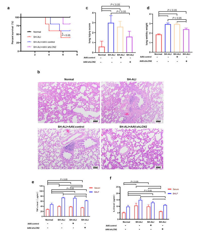
(a) Survival rate of rats in each group. Survival was monitored for 7 h, and survival curves were compared using log-rank test. Data are expressed as survival percentage. *P < 0.05 vs. SH-ALI + AAV.shLCN2 group. (b) Representative hematoxylin and eosin (H&E)-stained lung tissue sections from each group. (c) Lung injury scores in each group. (d) Lung wet-to-dry (W/D) weight ratios in each group; (e-f) Levels of inflammatory cytokines TNF-α (e) and IL-6 (f) in serum and BALF from each group. Data are expressed as means ± SD (n = 5). Abbreviations: LCN2, lipocalin-2; SH-ALI, severe hypothermia-induced acute lung injury; IL-6, interleukin-6; TNF-α, tumor necrosis factor-α; BALF, bronchoalveolar lavage fluid.

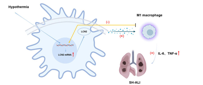
Figure 6. Schematic illustration of the promotive role of LCN2 in M1 polarization and SH-ALI
Hypothermic stimulation upregulates LCN2 expression in the lung. Elevated LCN2 promotes macrophage polarization toward the M1 phenotype, leading to increased release of pro-inflammatory mediators and amplification of the inflammatory response. This process ultimately contributes to the development of ALI and may progress to ARDS. Silencing of LCN2 attenuates lung injury by inhibiting M1 polarization and reducing inflammatory cytokine production in SH-ALI rats. Abbreviations: LCN2, lipocalin-2; ALI, acute lung injury; SH-ALI, severe hypothermia-induced acute lung injury; ARDS, acute respiratory distress syndrome. Symbols: (-) indicates inhibition; (+) indicates promotion.
Table 1. Primer sequences used for quantitative PCR
Target Forward Primer (5'-3') Reverse Primer (5'-3') Ldlr CATTTTCAGTGCCAACCGCC TGCCTCACACCAGTTTACCC Dhcr24 GCCCTTGGTGTCTATGGGTC CAGGATTAGCTCGTAGGCCG Cyp51 CCAGCCGAGGACATTCTTCA CCGCACACCGTTTTCTGTTC LCN2 GCTGTCGCTACTGGATCAGA TCGCTCCTTCAGTTCATCGG CCr7 TGTGTGACTTCAACTGCCCA GCACATCCTTCTTGAAGCAC Ccl2 CACTCACCTGCTGCTACTCA CCTTATTGGGGTCAGCACAGA Fabp4 ATGGTGGGGTCCTGGTACAT CTTGGCTCATGCCCTTTCGT Clec7a CATCCCAAACTACAGGCGTC ACTTGAAACGAGTTGGGGGT Acsl1 TGTGGGGTGGAAATCATCGG CATTGCTCCTTTGGGGTTGC Cybb TGCCAGTGTGTCGGAATCTC TGTGAATGGCCGTGTGAAGT Agt GCACGACTTCCTGACTTGG ACTCTGTGGGCTGCTCCTCC GAPDH CGTGTTCCTACCCCCAATGT AGGAGACAACCTGGTCCTCA  下载: 导出CSV
-
[1] Tomassini L, Lancia M, Gambelunghe C, et al. Immunohistochemical insights into hypothermia-related deaths: A systematic review. Forensic Sci Med Pathol, 2025; 21: 1358-1369. doi: 10.1007/s12024-024-00934-0 [2] Paal P, Pasquier M, Darocha T, et al. Accidental Hypothermia: 2021 Update. Int J Environ Res Public Health, 2022; 19(1): 501. doi: 10.3390/ijerph19010501 [3] Shahrbabak S M, Bouzid Z, Inan O T, et al. Physiology and enabling technologies for quantitative assessment of survivability during cold water immersion and rewarming: A review. Prog Biomed Eng (Bristol), 2025; 7(4): 1-23. doi: 10.1088/2516-1091/ae0c4b [4] Hleșcu A A, Grigoraș A, Ianole V, et al. Advanced diagnostic tools in hypothermia-related fatalities-a pathological perspective. Diagnostics (Basel), 2024; 14(7): 739. doi: 10.3390/diagnostics14070739 [5] Ding Y, Xu Y, Wang J, et al. Cold-inducible RNA-binding protein causes prolonged immersion-induced hypothermic acute lung injury in a rat model through TREM-1 and NF-κB. Faseb J, 2025; 39: e70955. doi: 10.1096/fj.202402540RRR [6] Liu J, Wu J, Qiao C, et al. Impact of chronic cold exposure on lung inflammation, pyroptosis and oxidative stress in mice. Int Immunopharmacol, 2023; 115: 109590. doi: 10.1016/j.intimp.2022.109590 [7] Shapiro K A, Phillips T C, Dineen E H, et al. The heart underwater: mechanisms and treatments of immersion pulmonary edema. Current Treatment Options in Cardiovascular Medicine, 2025; 19(27): 1075-1079. doi: 10.1007/s11936-025-01075-5 [8] Wang Z, Wang Z. The role of macrophages polarization in sepsis-induced acute lung injury. Front Immunol, 2023; 14: 1209438. doi: 10.3389/fimmu.2023.1209438 [9] Liu C, Xiao K, Xie L. Advances in the regulation of macrophage polarization by mesenchymal stem cells and implications for ALI/ARDS treatment. Front Immunol, 2022; 13: 928134. doi: 10.3389/fimmu.2022.928134 [10] Zhu G, Yu H, Peng T, et al. Glycolytic enzyme PGK1 promotes M1 macrophage polarization and induces pyroptosis of acute lung injury via regulation of NLRP3. Respir Res, 2024; 25: 291. doi: 10.1186/s12931-024-02926-8 [11] Yan M, Tang J, Liu Y, et al. Progress of alveolar macrophages in biological function and acute lung injury/acute respiratory distress syndrome. Front Immunol, 2025; 16: 1683411. doi: 10.3389/fimmu.2025.1683411 [12] Deshpande D, Chhugani K, Chang Y, et al. RNA-seq data science: From raw data to efective interpretation. Front Genet, 2023; 14: 997383. doi: 10.3389/fgene.2023.997383 [13] Pasmanik-Chor M. Biological perspectives of RNA-sequencing experimental design. Methods Mol Biol, 2021; 2243: 327-337. doi: 10.1007/978-1-0716-1103-6_17 [14] Cheng L, Xing H, Mao X, et al. Lipocalin-2 promotes m1 macrophages polarization in a mouse cardiac ischaemia-reperfusion injury model. Scand J Immunol, 2015; 81: 31-38. doi: 10.1111/sji.12245 [15] Reynolds P, Wall P, Van Griensven M, et al. Shock supports the use of animal research reporting guidelines. Shock, 2012; 38: 1-3. doi: 10.1097/SHK.0b013e31825f396c [16] Huang H, Wang J, Hussain S A, et al. Gossypin exert lipopolysaccharide induced lung inflammation via alteration of Nrf2/HO-1 and NF-κB signaling pathway. Environ Toxicol, 2023; 38: 1786-1799. doi: 10.1002/tox.23806 [17] Konkimalla A, Elmore Z, Konishi S, et al. Eficient Adeno-associated virus-mediated transgenesis in alveolar stem cells and associated niches. Am J Respir Cell Mol Biol, 2023; 69: 255-265. doi: 10.1165/rcmb.2022-0424MA [18] El Fakihi S, El Allam A, Tahoune H, et al. Functional characterization of small and large alveolar macrophages in sarcoidosis and idiopathic pulmonary fibrosis compared with non-fibrosis interstitial lung diseases. Hum Antibodies, 2023; 31: 59-69. doi: 10.3233/HAB-230005 [19] Smith K M, Mrozek J D, Simonton S C, et al. Prolonged partial liquid ventilation using conventional and high-frequency ventilatory techniques: gas exchange and lung pathology in an animal model of respiratory distress syndrome. Crit Care Med, 1997; 25: 1888-1897. doi: 10.1097/00003246-199711000-00030 [20] Tang J, Ding Y, Chen W, et al. VASP knockdown ameliorates lipopolysaccharide-induced acute lung injury with inhibition of M1 macrophage polarization through the cGMP-PKG signaling pathway. Inflammation, 2025; 48(5): 3458-3471. doi: 10.1007/s10753-025-02277-6 [21] Wiberg S, Mortensen A F, Kjaergaard J, et al. Accidental hypothermia in Denmark: A nationwide cohort study of incidence and outcomes. BMJ Open, 2021; 11: e046806. doi: 10.1136/bmjopen-2020-046806 [22] Dickinson G M, Maya G X, Lo Y, et al. Hypothermia-related deaths: a 10-year retrospective study of two major metropolitan cities in the United States. J Forensic Sci, 2020; 65: 2013-2018. doi: 10.1111/1556-4029.14518 [23] Bjertnæs L J, Næsheim T O, Reierth E, et al. Physiological changes in subjects exposed to accidental hypothermia: An update. Front Med (Lausanne), 2022; 9: 824395. doi: 10.3389/fmed.2022.824395 [24] Kulkarni H S, Lee J S, Bastarache J A, et al. Update on the features and measurements of experimental acute lung injury in animals: an oficial American thoracic society workshop report. Am J Respir Cell Mol Biol, 2022; 66: e1-e14. [25] Chen X, Tang J, Shuai W, et al. Macrophage polarization and its role in the pathogenesis of acute lung injury/acute respiratory distress syndrome. Inflamm Res, 2020; 69: 883-895. doi: 10.1007/s00011-020-01378-2 [26] Shan Q, Dong Z, Li N, et al. Deciphering the heterogeneity of pulmonary macrophages in response to fine particles. Small, 2026; 22: e07293. doi: 10.1002/smll.202507293 [27] Speth J M, Bourdonnay E, Penke L R, et al. Alveolar epithelial cell-derived prostaglandin E2 serves as a request signal for macrophage secretion of suppressor of cytokine signaling 3 during innate inflammation. J Immunol, 2016; 196: 5112-5120. doi: 10.4049/jimmunol.1502153 [28] Morrison T J, Jackson M V, Cunningham E K, et al. Mesenchymal stromal cells modulate macrophages in clinically relevant lung injury models by extracellular vesicle mitochondrial transfer. Am J Respir Crit Care Med, 2017; 196: 1275-1286. doi: 10.1164/rccm.201701-0170OC [29] Guardado S, Ojeda-Juárez D, Kaul M, et al. Comprehensive review of lipocalin 2-mediated efects in lung inflammation. Am J Physiol Lung Cell Mol Physiol, 2021; 321: L726-L733. doi: 10.1152/ajplung.00080.2021 [30] Sciarretta F, Ceci V, Tiberi M, et al. Lipocalin-2 promotes adipose-macrophage interactions to shape peripheral and central inflammatory responses in experimental autoimmune encephalomyelitis. Mol Metab, 2023; 76: 101783. doi: 10.1016/j.molmet.2023.101783 [31] An H S, Lee J, Lee S J, et al. Lipocalin-2 deletion attenuates lipopolysaccharide-induced acute lung inflammation via downregulating chemotaxis-related genes. Biochem Biophys Res Commun, 2023; 652: 14-21. doi: 10.1016/j.bbrc.2023.02.029 [32] An H S, Yoo J W, Jeong J H, et al. Lipocalin-2 promotes acute lung inflammation and oxidative stress by enhancing macrophage iron accumulation. Int J Biol Sci, 2023; 19: 1163-1177. doi: 10.7150/ijbs.79915 [33] Wang X, Zhang C, Zou N, et al. Lipocalin-2 silencing suppresses inflammation and oxidative stress of acute respiratory distress syndrome by ferroptosis via inhibition of MAPK/ERK pathway in neonatal mice. Bioengineered, 2022; 13: 508-520. doi: 10.1080/21655979.2021.2009970 [34] Lu F, Inoue K, Kato J, et al. Functions and regulation of lipocalin-2 in gut-origin sepsis: A narrative review. Crit Care, 2019; 23: 269. doi: 10.1186/s13054-019-2550-2 [35] Du H, Liang L, Li J, et al. Lipocalin-2 alleviates LPS-induced inflammation through alteration of macrophage properties. J Inflamm Res, 2021; 14: 4189-4203. doi: 10.2147/JIR.S328916 [36] Lindstrom E, Deis J, Bernlohr D A, et al. Lipocalin 2 in obesity and diabetes: insights into its role in energy metabolism. Endocrines, 2025; 6(1): 4. doi: 10.3390/endocrines6010004 [37] Jensen B L. Beyond being a biomarker: lipocalin-2/NGAL as a facilitator for protective drug action in hypoxic kidney injury. Acta Physiol (Oxf), 2025; 241: e70110. doi: 10.1111/apha.70110 [38] Zhang Z X, Peng J, Ding W W. Lipocalin-2 and intestinal diseases. World J Gastroenterol, 2024; 30: 4864-4879. doi: 10.3748/wjg.v30.i46.4864 [39] Mitsui Y, Satoh T. Functional diversity of disorder-specific macrophages involved in various diseases. Inflamm Regen, 2025; 45: 29. doi: 10.1186/s41232-025-00390-5 [40] Hume P S, Lyn-Kew K H, Wynn E A, et al. Spatial heterogeneity of macrophages in the human lung. (2025-05-30) [2026-03-02]. https://doi.org/10.1101/2025.05.30.657106 .[41] Chen X, Wang F, Tang J, et al. Paralemmin-3 augments lipopolysaccharide-induced acute lung injury with M1 macrophage polarization via the notch signaling pathway. Respir Physiol Neurobiol, 2024; 320: 104203. doi: 10.1016/j.resp.2023.104203 [42] Lv Y, Zhang L. IRF7 Activates LCN2 Transcription to enhance LPS-induced acute lung injury by inducing macrophage ferroptosis and M1 polarization. Cell Biochem Biophys, 2025; 83: 2415-2430. doi: 10.1007/s12013-024-01651-9 -

 点击查看大图
计量
- 文章访问数: 194
- HTML全文浏览量: 97
- PDF下载量: 2
- 被引次数: 0

