Bronchial inflammatory profile in interferon-gamma-mediated immune response in asthma patients during airway response to cold stimulus
doi: 10.2478/fzm-2022-0031
-
Abstract:
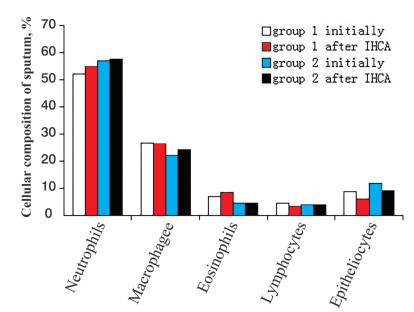
Objective To evaluate the inflammatory pattern and the interferon (IFN) -γ in the bronchial secretion of asthma patients in response to acute cold bronchoprovocation. Material and methods We enrolled 42 patients with asthma. We assessed asthma by Asthma Control Test, the lung function by spirometry before and after the bronchodilator test, followed by collecting induced sputum. The next day, we collected exhaled breath condensate (EBC) and conducted a 3-minute isocapnic hyperventilation with cold air (IHCA), followed by collecting spontaneously produced sputum. Results Group 1 included 20 patients with cold airway hyperresponsiveness (CAHR), and group 2 included 22 patients without CAHR. In both groups, a high level of neutrophils in bronchial secretion was observed before and after IHCA. In response to IHCA, the number of epitheliocytes in the sputum decreased to a greater extent in patients of group 1.The baseline epitheliocytes and the concentration of IFN-γ after IHCA had an inverse relationship (r = -0.60; P = 0.017). The baseline IFN-γ in EBC before and after IHCA was lower in group 1. Airway response to cold exposure directly correlated with IFN-γ levels after IHCA (Rs = 0.42; P = 0.014). Conclusion In asthma patients with CAHR, there is a relationship between the persistence of mixed inflammation and the level of IFN-γ in the bronchi. IFN-γ in response to IHCA is decreased with increased cytokine utilization during cold bronchospasm, which is accompanied by the mobilization of neutrophils and the shift in the cytokine spectrum of the respiratory tract towards the T helper cells (Th) 1 immune response. -
Table 1. Lung function and FEV1 dynamics after administration of salbutamol in asthma patients with different types of airway response to IHCA
Indicators Group 1 (n = 20) Group 2 (n = 22) P value predicted FVC, % 106.9 ± 2.1 110.3 ± 1.8 > 0.05 predicted FEV1, % 90.1 ± 2.1 98.0± 1.9 0.0019 FEV1/VC, % 70.3 ± 1.3 74.1± 1.1 0.0057 predicted MEF25-75, % 60.3 ± 3.0 70.7± 3.4 0.032 ∆FEV1bronchodilator, % 10.05 (4.00, 18.70) 7.28(3.37, 13.05) > 0.05 Values are presented as mean ± SE and median (Q1, Q3). IHCA, isocapnic hyperventilation with cold air; IFN, interferon; FVC, forced vital capacity; MEF25-75, the maximal midexpiratory flow at the level of 25%-75% FVC; ∆FEV1bronchodilator, %, change in the index after inhalation of a short-acting β2-agonist (salbutamol, 400 μg). Table 2. The concentration of IFN-γ in the exhaled breath condensate in asthma patients with different types of airway response to IHCA
Indicators Group 1 (n = 20) Group 2 (n = 22) P Value Initial 17.3± 2.8 21.2± 3.4 > 0.05 After IHCA 13.6± 3.2# 48.7± 5.4## 0.0024 Values are presented as mean ± SE. FVC, forced vital capacity; IHCA, isocapnic hyperventilation with cold air; IFN, interferon; P, significance level between groups 1 and 2 (unpaired t-test); #, P > 0.05; ##, P = 0.022. -
[1] Prikhodko A G, Perelman J M, Kolosov V P. Airway Hyperresponsiveness. Dal'nauka 2011. (In Russian) [2] PirogovA B, KolosovV P, PerelmanJ M, et al. Airway inflammation patterns and clinical and functional features in patients with severe uncontrolled asthma and cold-induced airway hyperresponsiveness. Pulmonologiya, 2016; 26(6): 701-707. (In Russian) [3] Hastie A T, Moore W C, Meyers D A, et al. Аnalyses of asthma severity phenotypes and inflammatory proteins in subjects stratified by sputum granulocytes. J Allergy Clin Immunol, 2010; 125(5): 1028-1036. doi: 10.1016/j.jaci.2010.02.008 [4] Petrova E S, Goryachev D V, Kuznetsova A D. Planning a clinical development programme for medicines for bronchial asthma. The Bulletin of the Scientific Centre for Expert Evaluation of Medicinal Products, 2021; 11(1): 55-69. (In Russian) doi: 10.30895/1991-2919-2021-11-1-55-69 [5] Duvall M G, Krishnamoorthy N, Levy B D. Non-type 2 inflammation in severe asthma is propelled by neutrophil cytoplasts and maintained by defective resolution. Allergol Int, 2019; 68(2): 143-149. doi: 10.1016/j.alit.2018.11.006 [6] Nikolskii A A, Shilovskiy I P, Yumashev K V, et al. Effect of local suppression of Stat3 gene expression in a mouse model of pulmonary neutrophilic inflammation. Immunology, 2021; 42(6): 600-614. (In Russian) doi: 10.33029/0206-4952-2021-42-6-600-614 [7] Lutckii A A, Zhirkov A A, Lobzin D Y, et al. Interferon-γ: biological function and application for study of cellular immune response. Journal Infectology, 2015; 7(4): 10-22. (In Russian) [8] Akira S. The role of IL-18 in innate immunity. Curr Opin Immunol, 2000; 12(1): 59-63. doi: 10.1016/S0952-7915(99)00051-5 [9] Hamza T, Barnett J B, Li B. Interleukin 12 a key immunoregulatory cytokine in infection applications. Int J Mol Sci, 2010; 11(3): 789-806. doi: 10.3390/ijms11030789 [10] Schroder K, Hertzog P J, Ravasi T, et al. Interferon-gamma: an overview of signals, mechanisms and functions. J Leukoc Biol, 2004; 75(2): 163-189. doi: 10.1189/jlb.0603252 [11] Gattoni A, Parlato A, Vangieri B, et al. Interferon-gamma: biologic functions and HCV therapy (type Ⅰ/Ⅱ) (1 of 2 parts). Clin Ter, 2006; 157(4): 377-386. [12] Global Initiative for Asthma (GINA). Global strategy for asthma management and prevention (2021 update). http://www.ginasthma.org. Last accessed on April 05, 2022. [13] Bakakos P, Schleich F, Alchanatis M, et al. Induced sputum in asthma: From bench to bedside. Curr Med Chem, 2011; 18 (10): 1415-1422. doi: 10.2174/092986711795328337 [14] Graham B L, Steenbruggen I, Miller M R, et al. Standardization of Spirometry 2019 Update. An Official American Thoracic Society and European Respiratory Society Technical Statement. Am J Respir Crit Care Med, 2019; 200(8): e70-e88. doi: 10.1164/rccm.201908-1590ST [15] Ульянычев Н. В. Системность научных исследований в медицине. Saarbrücken: LAP LAMBERT, 2014; 140 с. [16] Ray A, Kolls J K. Neutrophilic inflammation in asthma and association with disease severity. Trends Immunol, 2017; 38(12): 942-954. doi: 10.1016/j.it.2017.07.003 [17] Mall M A. Role of cilia, mucus, and airway surface liquid in mucociliary dysfunction: lessons from mouse models. J Aerosol Med Pulm Drug Deliv, 2008; 21(1): 13-24. doi: 10.1089/jamp.2007.0659 [18] Pirogov A B, Zinov'ev S V, Prikhodko А G, et al. Features of structural organization of goblet epithelium of bronchi in asthma patients with cold airway hyperresponsiveness. Bulletin Physiology and Pathology of Respiration, 2018; 67: 17-24. (In Russian) [19] Pirogov A B, Prikhodko А G, Perelman J M, et al. Profile of bronchial inflammation and clinical features of mild bronchial asthma. Bulletin Physiology and Pathology of Respiration, 2018; 70: 8-14. (In Russian) [20] Shuai K, Liu B. Regulation of JAK-STAT signalling in the immune system. Nat Rev Immunol, 2003; 3(11): 942-954. [21] Mineev V N, Sorokina L N, Nyoma M A. Effects of IL-4 upon the activity of stat6 transcription factor in peripheral blood lymphocytes in bronchial asthma. Medical Immunology (Russia), 2009; 11(2-3): 177-184. (In Russian) [22] Kiwamoto T, Ishii Y, Morishima Y, et al. Transcription factors T-bet and GATA-3 regulate development of airway remodeling. Am J Respir Crit Care Med, 2006; 174(2): 142-151. doi: 10.1164/rccm.200601-079OC [23] Usui T, Preiss J C, Kanno Y, et al. T-bet regulates Th1 responses through essential effects on GATA-3 function rather than on IFNG gene acetylation and transcription. J Exp Med, 2006; 203(3): 755-766. doi: 10.1084/jem.20052165 [24] Zhu J, Yamane H, Cote-Sierra J, et al. GATA-3 promotes Th2 responses through three different mechanisms: induction of Th2 cytokine production, selective growth of Th2 cells and inhibition of Th1 cell-specific factors. Cell Res, 2006; 16(1): 3-10. doi: 10.1038/sj.cr.7310002 [25] Mineev V N, Sorokina L N, Nyoma M A, et al. GATA-3 expression in peripheral blood lymphocytes of patients with bronchial asthma. Medical Immunology (Russia), 2010; 12(1-2): 21-28. (In Russian) [26] Kulikov E S, Ogorodova L M, Freidin M B, et al. Molecular mechanisms of severe asthma. Mol Med, 2013; 2: 24-32. (In Russian) [27] Zinatizadeha M R, Schockb B, Chalbatani G M, et al. The Nuclear Factor Kappa B (NF-κB) signaling in cancer development and immune diseases. Genes Dis, 2021; 8(3): 287-297. [28] Mayansky A N, Mayansky N A, Zaslavskaya M I. Nuclear factor kappa B and inflammation. Cytokines and inflammation, 2007; 6(2): 3-9. (In Russian) -


 投稿系统
投稿系统


 下载:
下载:


